
-retinoic acid</span>;</li> <li class='image_legend_li'>1 copy of <span class='highlight'>water</span>.</li></ul>")
-retinoic acid</span>;</li> <li class='image_legend_li'>1 copy of <span class='highlight'>water</span>.</li></ul>")
-retinoic acid</span>;</li> <li class='image_legend_li'>1 copy of <span class='highlight'>water</span>.</li></ul>")
Function and Biology Details
Biochemical function:
Biological process:
Cellular component:
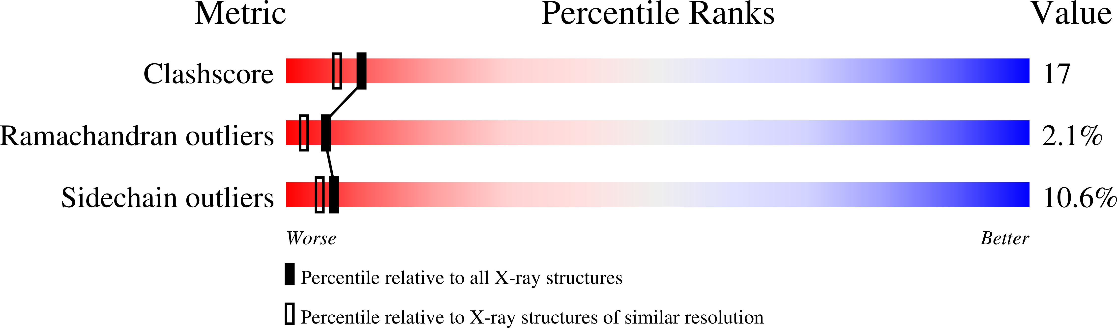
Structure analysis Details
Assembly composition:
homo dimer (preferred)
Assembly name:
RXRalpha-RXRalpha retinoic acid receptor complex (preferred)
PDBe Complex ID:
PDB-CPX-148719 (preferred)
Entry contents:
1 distinct polypeptide molecule
Macromolecule:
Retinoic acid receptor RXR-alpha
Molecule details ›
Chains: A, B
Length: 239 amino acids
Theoretical weight: 26.75 KDa
Source organism: Homo sapiens
Expression system: Escherichia coli
UniProt:
Sequence domains: Ligand-binding domain of nuclear hormone receptor
Structure domains: Retinoid X Receptor
Length: 239 amino acids
Theoretical weight: 26.75 KDa
Source organism: Homo sapiens
Expression system: Escherichia coli
UniProt:
- Canonical:
 P19793 (Residues: 224-462; Coverage: 52%)
Sequence domains: Ligand-binding domain of nuclear hormone receptor
Structure domains: Retinoid X Receptor

