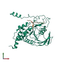
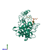
1fpr

X-ray diffraction
2.5Å resolution

CRYSTAL STRUCTURE OF THE COMPLEX FORMED BETWEEN THE CATALYTIC DOMAIN OF SHP-1 AND AN IN VITRO PEPTIDE SUBSTRATE PY469 DERIVED FROM SHPS-1.

Released:
Source organism: Homo sapiens

Function and Biology Details

Reaction catalysed:
Protein tyrosine phosphate + H(2)O = protein tyrosine + phosphate
Biological process:
Cellular component:
  • not assigned

Structure analysis Details

Assemblies composition:
hetero dimer (preferred)
hetero tetramer
PDBe Complex ID:
PDB-CPX-151412 (preferred)
Entry contents:
2 distinct polypeptide molecules
Macromolecules (2 distinct):
Tyrosine-protein phosphatase non-receptor type 6 Chain: A
Molecule details ›
Chain: A
Length: 284 amino acids
Theoretical weight: 32.56 KDa
Source organism: Homo sapiens
Expression system: Escherichia coli
UniProt:
  • Canonical: P29350 (Residues: 243-526; Coverage: 48%)
Gene names: HCP, PTP1C, PTPN6
Sequence domains: Protein-tyrosine phosphatase
Structure domains: Protein tyrosine phosphatase superfamily
PEPTIDE PY469 Chain: B
Molecule details ›
Chain: B
Length: 10 amino acids
Theoretical weight: 1.24 KDa
Source organism: Homo sapiens
Expression system: Not provided

Ligands and Environments

No bound ligands
1 modified residue:

Experiments and Validation Details

Entry percentile scores
X-ray source: RIGAKU RU300
Spacegroup: P21212
Unit cell:
a: 111.58Å b: 45.21Å c: 56.27Å
α: 90° β: 90° γ: 90°
R-values:
R R work R free
0.199 0.194 0.303
Expression systems:
  • Escherichia coli
  • Not provided