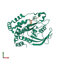
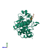
1g1g

X-ray diffraction
2.2Å resolution

CRYSTAL STRUCTURE OF PROTEIN TYROSINE PHOSPHATASE 1B COMPLEXED WITH A MONO-PHOSPHORYLATED PEPTIDE (ETDY(PTR)RKGGKGLL) FROM THE INSULIN RECEPTOR KINASE

Released:

Function and Biology Details

Reaction catalysed:
Protein tyrosine phosphate + H(2)O = protein tyrosine + phosphate
Biological process:
Cellular component:
  • not assigned

Structure analysis Details

Assembly composition:
hetero dimer (preferred)
PDBe Complex ID:
PDB-CPX-148264 (preferred)
Entry contents:
2 distinct polypeptide molecules
Macromolecules (2 distinct):
Tyrosine-protein phosphatase non-receptor type 1 Chain: A
Molecule details ›
Chain: A
Length: 298 amino acids
Theoretical weight: 34.69 KDa
Source organism: Homo sapiens
Expression system: Escherichia coli
UniProt:
  • Canonical: P18031 (Residues: 1-298; Coverage: 69%)
Gene names: PTP1B, PTPN1
Sequence domains: Protein-tyrosine phosphatase
Structure domains: Protein tyrosine phosphatase superfamily
MONO-PHOSPHORYLATED PEPTIDE FROM THE INSULIN RECEPTOR KINASE Chain: B
Molecule details ›
Chain: B
Length: 13 amino acids
Theoretical weight: 1.58 KDa
Source organism: Homo sapiens
Expression system: Not provided

Ligands and Environments

No bound ligands
1 modified residue:

Experiments and Validation Details

Entry percentile scores
X-ray source: SRS BEAMLINE PX9.6
Spacegroup: P21
Unit cell:
a: 44.492Å b: 89.281Å c: 52.922Å
α: 90° β: 99.73° γ: 90°
R-values:
R R work R free
0.212 0.212 0.228
Expression systems:
  • Escherichia coli
  • Not provided