1iod

X-ray diffraction
2.3Å resolution

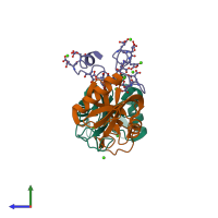
CRYSTAL STRUCTURE OF THE COMPLEX BETWEEN THE COAGULATION FACTOR X BINDING PROTEIN FROM SNAKE VENOM AND THE GLA DOMAIN OF FACTOR X

Released:
Primary publication:
Crystal structure of an anticoagulant protein in complex with the Gla domain of factor X.
Proc Natl Acad Sci U S A 98 7230-4 (2001)
PMID: 11404471

Function and Biology Details

Structure analysis Details

Assemblies composition:
hetero trimer (preferred)
hetero hexamer
PDBe Complex ID:
PDB-CPX-133222 (preferred)
Entry contents:
3 distinct polypeptide molecules
Macromolecules (3 distinct):
Snaclec agkisacutacin subunit A Chain: A
Molecule details ›
Chain: A
Length: 129 amino acids
Theoretical weight: 14.68 KDa
Source organism: Deinagkistrodon acutus
UniProt:
  • Canonical: Q9IAM1 (Residues: 24-152; Coverage: 100%)
Sequence domains: Lectin C-type domain
Structure domains: Mannose-Binding Protein A, subunit A
Snaclec anticoagulant protein subunit B Chain: B
Molecule details ›
Chain: B
Length: 123 amino acids
Theoretical weight: 14.58 KDa
Source organism: Deinagkistrodon acutus
UniProt:
  • Canonical: Q9DEF8 (Residues: 24-146; Coverage: 100%)
Sequence domains: Lectin C-type domain
Structure domains: Mannose-Binding Protein A, subunit A
Factor X light chain Chain: G
Molecule details ›
Chain: G
Length: 44 amino acids
Theoretical weight: 5.73 KDa
Source organism: Bos taurus
UniProt:
  • Canonical: P00743 (Residues: 41-84; Coverage: 9%)
Gene name: F10
Sequence domains: Vitamin K-dependent carboxylation/gamma-carboxyglutamic (GLA) domain

Ligands and Environments

1 bound ligand:
1 modified residue:

Experiments and Validation Details

Entry percentile scores
X-ray source: PHOTON FACTORY BEAMLINE BL-6A
Spacegroup: P41212
Unit cell:
a: 99.76Å b: 99.76Å c: 90.42Å
α: 90° β: 90° γ: 90°
R-values:
R R work R free
0.225 0.201 0.267