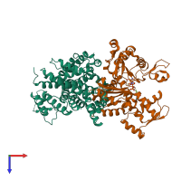
1ma9

X-ray diffraction
2.4Å resolution

Crystal structure of the complex of human vitamin D binding protein and rabbit muscle actin

Released:
Primary publication:
Actin-DBP: the perfect structural fit?
Acta Crystallogr D Biol Crystallogr 59 263-73 (2003)
PMID: 12554937

Function and Biology Details

Structure analysis Details

Assembly composition:
hetero dimer (preferred)
PDBe Complex ID:
PDB-CPX-136196 (preferred)
Entry contents:
2 distinct polypeptide molecules
Macromolecules (2 distinct):
Vitamin D-binding protein Chain: A
Molecule details ›
Chain: A
Length: 458 amino acids
Theoretical weight: 51.29 KDa
Source organism: Homo sapiens
UniProt:
  • Canonical: P02774 (Residues: 17-474; Coverage: 100%)
Gene name: GC
Sequence domains:
Structure domains: Serum Albumin; Chain A, Domain 1
Actin, alpha skeletal muscle Chain: B
Molecule details ›
Chain: B
Length: 375 amino acids
Theoretical weight: 41.88 KDa
Source organism: Oryctolagus cuniculus
UniProt:
  • Canonical: P68135 (Residues: 3-377; Coverage: 100%)
Gene names: ACTA, ACTA1
Sequence domains: Actin
Structure domains:

Ligands and Environments

2 bound ligands:
1 modified residue:

Experiments and Validation Details

Entry percentile scores
X-ray source: EMBL/DESY, HAMBURG BEAMLINE BW7B
Spacegroup: P21
Unit cell:
a: 74.44Å b: 74.9Å c: 88.02Å
α: 90° β: 110.19° γ: 90°
R-values:
R R work R free
0.203 0.2 0.25