1owr

X-ray diffraction
3Å resolution

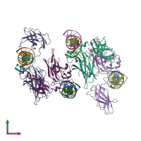
CRYSTAL STRUCTURE OF HUMAN NFAT1 BOUND MONOMERICALLY TO DNA

Released:
Source organism: Homo sapiens
Primary publication:
Structure of NFAT bound to DNA as a monomer.
J Mol Biol 334 1009-22 (2003)
PMID: 14643663

Function and Biology Details

Structure analysis Details

Assembly composition:
hetero trimer (preferred)
PDBe Complex ID:
PDB-CPX-114333 (preferred)
Entry contents:
1 distinct polypeptide molecule
2 distinct DNA molecules
Macromolecules (3 distinct):
Nuclear factor of activated T-cells, cytoplasmic 2 Chains: M, N, P, Q
Molecule details ›
Chains: M, N, P, Q
Length: 284 amino acids
Theoretical weight: 32.31 KDa
Source organism: Homo sapiens
Expression system: Escherichia coli
UniProt:
  • Canonical: Q13469 (Residues: 396-678; Coverage: 31%)
Gene names: NFAT1, NFATC2, NFATP
Sequence domains:
Structure domains:
NFAT1 Monomeric Binding Site, Plus Strand Chains: A, C, E, G
Molecule details ›
Chains: A, C, E, G
Length: 15 nucleotides
Theoretical weight: 4.66 KDa
Source organism: Homo sapiens
Expression system: Not provided
NFAT1 Monomeric Binding Site, Minus Strand Chains: B, D, F, H
Molecule details ›
Chains: B, D, F, H
Length: 15 nucleotides
Theoretical weight: 4.52 KDa
Source organism: Homo sapiens
Expression system: Not provided

Ligands and Environments

No bound ligands
No modified residues

Experiments and Validation Details

Entry percentile scores
X-ray source: APS BEAMLINE 14-BM-C
Spacegroup: P21
Unit cell:
a: 100.646Å b: 94.849Å c: 112.788Å
α: 90° β: 104.34° γ: 90°
R-values:
R R work R free
0.241 0.241 0.273
Expression systems:
  • Escherichia coli
  • Not provided