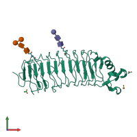
1ozn

X-ray diffraction
1.52Å resolution

1.5A Crystal Structure of the Nogo Receptor Ligand Binding Domain Reveals a Convergent Recognition Scaffold Mediating Inhibition of Myelination

Released:

Function and Biology Details

Biochemical function:
  • not assigned
Biological process:
  • not assigned
Cellular component:
  • not assigned

Structure analysis Details

Assembly composition:
monomeric (preferred)
Assembly name:
PDBe Complex ID:
PDB-CPX-189865 (preferred)
Entry contents:
1 distinct polypeptide molecule
Macromolecules (3 distinct):
Reticulon-4 receptor Chain: A
Molecule details ›
Chain: A
Length: 285 amino acids
Theoretical weight: 31.61 KDa
Source organism: Homo sapiens
Expression system: Trichoplusia ni
UniProt:
  • Canonical: Q9BZR6 (Residues: 26-310; Coverage: 64%)
Gene names: NOGOR, RTN4R, UNQ330/PRO526
Sequence domains: Leucine rich repeat
Structure domains: Ribonuclease Inhibitor

Ligands and Environments

Carbohydrate polymer : NEW Components: NAG, NDG, MAN
Carbohydrate polymer : NEW Components: NAG, NDG, BMA
1 bound ligand:
No modified residues

Experiments and Validation Details

Entry percentile scores
X-ray source: ALS BEAMLINE 8.2.1
Spacegroup: P1
Unit cell:
a: 32.206Å b: 33.915Å c: 60.221Å
α: 85.02° β: 75.91° γ: 67.96°
R-values:
R R work R free
0.168 0.167 0.198
Expression system: Trichoplusia ni