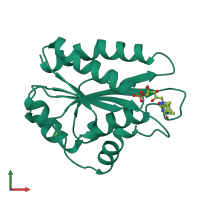
1b1c

X-ray diffraction
1.93Å resolution

CRYSTAL STRUCTURE OF THE FMN-BINDING DOMAIN OF HUMAN CYTOCHROME P450 REDUCTASE AT 1.93A RESOLUTION

Released:

Function and Biology Details

Reaction catalysed:
NADPH + n oxidized hemoprotein = NADP(+) + n reduced hemoprotein
Biochemical function:
Biological process:
  • not assigned
Cellular component:
  • not assigned

Structure analysis Details

Assembly composition:
monomeric (preferred)
PDBe Complex ID:
PDB-CPX-147724 (preferred)
Entry contents:
1 distinct polypeptide molecule
Macromolecule:
NADPH--cytochrome P450 reductase Chain: A
Molecule details ›
Chain: A
Length: 181 amino acids
Theoretical weight: 20.38 KDa
Source organism: Homo sapiens
UniProt:
  • Canonical: P16435 (Residues: 61-241; Coverage: 27%)
Gene names: CYPOR, POR
Sequence domains: Flavodoxin
Structure domains: Rossmann fold

Ligands and Environments


Cofactor: Ligand FMN 1 x FMN
1 bound ligand:
No modified residues

Experiments and Validation Details

Entry percentile scores
X-ray source: SIEMENS
Spacegroup: P21
Unit cell:
a: 39.31Å b: 51.44Å c: 47.6Å
α: 90° β: 105.93° γ: 90°
R-values:
R R work R free
0.197 0.197 0.243