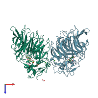
1c9u

X-ray diffraction
2.2Å resolution

CRYSTAL STRUCTURE OF THE SOLUBLE QUINOPROTEIN GLUCOSE DEHYDROGENASE IN COMPLEX WITH PQQ

Released:

Function and Biology Details

Reaction catalysed:
D-glucose + ubiquinone = D-glucono-1,5-lactone + ubiquinol
Biochemical function:
Biological process:
  • not assigned
Cellular component:
  • not assigned

Structure analysis Details

Assembly composition:
homo dimer (preferred)
PDBe Complex ID:
PDB-CPX-146684 (preferred)
Entry contents:
1 distinct polypeptide molecule
Macromolecule:
Quinoprotein glucose dehydrogenase B Chains: A, B
Molecule details ›
Chains: A, B
Length: 454 amino acids
Theoretical weight: 50.29 KDa
Source organism: Acinetobacter calcoaceticus
Expression system: Escherichia coli
UniProt:
  • Canonical: P13650 (Residues: 25-478; Coverage: 100%)
Gene name: gdhB
Sequence domains: Glucose / Sorbosone dehydrogenase
Structure domains: TolB, C-terminal domain

Ligands and Environments


Cofactor: Ligand PQQ 2 x PQQ
2 bound ligands:
No modified residues

Experiments and Validation Details

Entry percentile scores
X-ray source: ESRF BEAMLINE BM02
Spacegroup: C2221
Unit cell:
a: 60.59Å b: 158.72Å c: 221.39Å
α: 90° β: 90° γ: 90°
R-values:
R R work R free
0.224 0.223 0.286
Expression system: Escherichia coli