1ee6

X-ray diffraction
2.3Å resolution

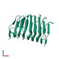
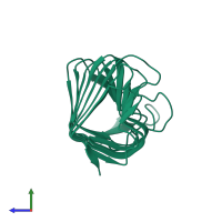
CRYSTAL STRUCTURE OF PECTATE LYASE FROM BACILLUS SP. STRAIN KSM-P15.

Released:
Source organism: Bacillus sp. KSM-P15
Primary publication:
The first structure of pectate lyase belonging to polysaccharide lyase family 3.
Acta Crystallogr D Biol Crystallogr 57 1786-92 (2001)
PMID: 11717490

Function and Biology Details

Reaction catalysed:
Eliminative cleavage of (1->4)-alpha-D-galacturonan to give oligosaccharides with 4-deoxy-alpha-D-galact-4-enuronosyl groups at their non-reducing ends
Biochemical function:
Biological process:
Cellular component:

Structure analysis Details

Assembly composition:
monomeric (preferred)
Assembly name:
PDBe Complex ID:
PDB-CPX-193177 (preferred)
Entry contents:
1 distinct polypeptide molecule
Macromolecule:
Pectate lyase Chain: A
Molecule details ›
Chain: A
Length: 197 amino acids
Theoretical weight: 20.95 KDa
Source organism: Bacillus sp. KSM-P15
UniProt:
  • Canonical: Q9RHW0 (Residues: 28-224; Coverage: 100%)
Sequence domains: Pectate lyase
Structure domains: Single-stranded right-handed beta-helix, Pectin lyase-like

Ligands and Environments

1 bound ligand:
No modified residues

Experiments and Validation Details

Entry percentile scores
X-ray source: RIGAKU RU300
Spacegroup: P212121
Unit cell:
a: 42.9Å b: 60.5Å c: 83Å
α: 90° β: 90° γ: 90°
R-values:
R R work R free
0.186 0.186 0.234