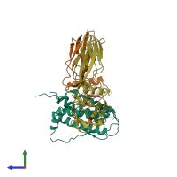
1eer

X-ray diffraction
1.9Å resolution

CRYSTAL STRUCTURE OF HUMAN ERYTHROPOIETIN COMPLEXED TO ITS RECEPTOR AT 1.9 ANGSTROMS

Released:

Function and Biology Details

Structure analysis Details

Assembly composition:
hetero trimer (preferred)
PDBe Complex ID:
PDB-CPX-134423 (preferred)
Entry contents:
2 distinct polypeptide molecules
Macromolecules (2 distinct):
Erythropoietin Chain: A
Molecule details ›
Chain: A
Length: 166 amino acids
Theoretical weight: 18.47 KDa
Source organism: Homo sapiens
Expression system: Escherichia coli
UniProt:
  • Canonical: P01588 (Residues: 28-193; Coverage: 100%)
Gene name: EPO
Sequence domains: Erythropoietin/thrombopoietin
Structure domains: Growth Hormone; Chain: A;
Erythropoietin receptor Chains: B, C
Molecule details ›
Chains: B, C
Length: 227 amino acids
Theoretical weight: 25.19 KDa
Source organism: Homo sapiens
Expression system: Komagataella pastoris
UniProt:
  • Canonical: P19235 (Residues: 26-250; Coverage: 46%)
Gene name: EPOR
Sequence domains:
Structure domains: Immunoglobulins

Ligands and Environments

No bound ligands
No modified residues

Experiments and Validation Details

Entry percentile scores
X-ray source: RIGAKU RUH3R
Spacegroup: P212121
Unit cell:
a: 58.4Å b: 79.3Å c: 136.5Å
α: 90° β: 90° γ: 90°
R-values:
R R work R free
0.242 0.242 0.318
Expression systems:
  • Escherichia coli
  • Komagataella pastoris