1fgg

X-ray diffraction
2.3Å resolution

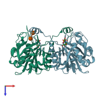
CRYSTAL STRUCTURE OF 1,3-GLUCURONYLTRANSFERASE I (GLCAT-I) COMPLEXED WITH GAL-GAL-XYL, UDP, AND MN2+

Released:

Function and Biology Details

Reaction catalysed:
UDP-alpha-D-glucuronate + [protein]-3-O-(beta-D-galactosyl-(1->3)-beta-D-galactosyl-(1->4)-beta-D-xylosyl)-L-serine = UDP + [protein]-3-O-(beta-D-GlcA-(1->3)-beta-D-Gal-(1->3)-beta-D-Gal-(1->4)-beta-D-Xyl)-L-serine
Biological process:
  • not assigned
Cellular component:

Structure analysis Details

Assembly composition:
homo dimer (preferred)
PDBe Complex ID:
PDB-CPX-131751 (preferred)
Entry contents:
1 distinct polypeptide molecule
Macromolecules (2 distinct):
Galactosylgalactosylxylosylprotein 3-beta-glucuronosyltransferase 3 Chains: A, B
Molecule details ›
Chains: A, B
Length: 261 amino acids
Theoretical weight: 28.99 KDa
Source organism: Homo sapiens
Expression system: Escherichia coli
UniProt:
  • Canonical: O94766 (Residues: 76-335; Coverage: 78%)
Gene name: B3GAT3
Sequence domains: Glycosyltransferase family 43
Structure domains: Spore Coat Polysaccharide Biosynthesis Protein SpsA; Chain A

Ligands and Environments

Carbohydrate polymer : NEW Components: GAL
3 bound ligands:
No modified residues

Experiments and Validation Details

Entry percentile scores
X-ray source: RIGAKU RU300
Spacegroup: P21
Unit cell:
a: 57.842Å b: 48.219Å c: 102.204Å
α: 90° β: 92.79° γ: 90°
R-values:
R R work R free
0.189 0.188 0.215
Expression system: Escherichia coli