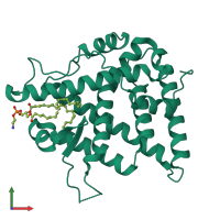
1g2n

X-ray diffraction
1.65Å resolution

CRYSTAL STRUCTURE OF THE LIGAND BINDING DOMAIN OF THE ULTRASPIRACLE PROTEIN USP, THE ORTHOLOG OF RXRS IN INSECTS

Released:

Function and Biology Details

Structure analysis Details

Assembly composition:
monomeric (preferred)
PDBe Complex ID:
PDB-CPX-181973 (preferred)
Entry contents:
1 distinct polypeptide molecule
Macromolecule:
NR LBD domain-containing protein Chain: A
Molecule details ›
Chain: A
Length: 264 amino acids
Theoretical weight: 29.96 KDa
Source organism: Heliothis virescens
Expression system: Escherichia coli BL21(DE3)
UniProt:
  • Canonical: Q7SIF6 (Residues: 1-264; Coverage: 100%)
Sequence domains: Ligand-binding domain of nuclear hormone receptor
Structure domains: Retinoid X Receptor

Ligands and Environments

1 bound ligand:
No modified residues

Experiments and Validation Details

Entry percentile scores
X-ray source: ESRF BEAMLINE ID14-2
Spacegroup: P4322
Unit cell:
a: 58.211Å b: 58.211Å c: 144.687Å
α: 90° β: 90° γ: 90°
R-values:
R R work R free
0.21 0.21 0.244
Expression system: Escherichia coli BL21(DE3)