1j5o

X-ray diffraction
3.5Å resolution

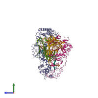
CRYSTAL STRUCTURE OF MET184ILE MUTANT OF HIV-1 REVERSE TRANSCRIPTASE IN COMPLEX WITH DOUBLE STRANDED DNA TEMPLATE-PRIMER

Released:

Function and Biology Details

Reactions catalysed:
Deoxynucleoside triphosphate + DNA(n) = diphosphate + DNA(n+1)
3'-end directed exonucleolytic cleavage of viral RNA-DNA hybrid
Endohydrolysis of RNA in RNA/DNA hybrids. Three different cleavage modes: 1. sequence-specific internal cleavage of RNA. Human immunodeficiency virus type 1 and Moloney murine leukemia virus enzymes prefer to cleave the RNA strand one nucleotide away from the RNA-DNA junction. 2. RNA 5'-end directed cleavage 13-19 nucleotides from the RNA end. 3. DNA 3'-end directed cleavage 15-20 nucleotides away from the primer terminus.
Specific for a P1 residue that is hydrophobic, and P1' variable, but often Pro.
Cellular component:
  • not assigned

Structure analysis Details

Assembly composition:
hetero hexamer (preferred)
PDBe Complex ID:
PDB-CPX-207778 (preferred)
Entry contents:
4 distinct polypeptide molecules
2 distinct DNA molecules
Macromolecules (6 distinct):
Reverse transcriptase/ribonuclease H Chain: A
Molecule details ›
Chain: A
Length: 558 amino acids
Theoretical weight: 64.26 KDa
Source organism: Human immunodeficiency virus 1
Expression system: Escherichia coli
UniProt:
  • Canonical: P03366 (Residues: 600-1157; Coverage: 39%)
Gene name: gag-pol
Sequence domains:
Structure domains:
p51 RT Chain: B
Molecule details ›
Chain: B
Length: 430 amino acids
Theoretical weight: 50.26 KDa
Source organism: Human immunodeficiency virus 1
Expression system: Escherichia coli
UniProt:
  • Canonical: P03366 (Residues: 600-1029; Coverage: 30%)
Gene name: gag-pol
Sequence domains:
Structure domains:
ANTIBODY (LIGHT CHAIN) Chain: L
Molecule details ›
Chain: L
Length: 214 amino acids
Theoretical weight: 22.98 KDa
Source organism: Mus musculus
Structure domains: Immunoglobulins
ANTIBODY (HEAVY CHAIN) Chain: H
Molecule details ›
Chain: H
Length: 220 amino acids
Theoretical weight: 23.46 KDa
Source organism: Mus musculus
Structure domains: Immunoglobulins
5'-D(*AP*TP*GP*GP*CP*GP*CP*CP*CP*GP*AP*AP*CP*AP*GP*GP*GP*AP*C)-3' Chain: T
Molecule details ›
Chain: T
Length: 19 nucleotides
Theoretical weight: 5.86 KDa
5'-D(*GP*TP*CP*CP*CP*TP*GP*TP*TP*CP*GP*GP*GP*CP*GP*CP*CP*A)-3' Chain: P
Molecule details ›
Chain: P
Length: 18 nucleotides
Theoretical weight: 5.48 KDa

Ligands and Environments

No bound ligands
No modified residues

Experiments and Validation Details

Entry percentile scores
X-ray source: CHESS BEAMLINE F1, NSLS BEAMLINE X25
Spacegroup: P3212
Unit cell:
a: 169.18Å b: 169.18Å c: 221.91Å
α: 90° β: 90° γ: 120°
R-values:
R R work R free
0.27 0.262 0.338
Expression system: Escherichia coli