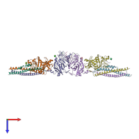
1n73

X-ray diffraction
2.9Å resolution

Fibrin D-Dimer, Lamprey complexed with the PEPTIDE LIGAND: GLY-HIS-ARG-PRO-AMIDE

Released:

Function and Biology Details

Structure analysis Details

Assembly composition:
hetero decamer (preferred)
PDBe Complex ID:
PDB-CPX-136033 (preferred)
Entry contents:
4 distinct polypeptide molecules
Macromolecules (4 distinct):
Fibrinogen alpha-1 chain Chains: A, D
Molecule details ›
Chains: A, D
Length: 119 amino acids
Theoretical weight: 14.07 KDa
Source organism: Petromyzon marinus
UniProt:
  • Canonical: P02674 (Residues: 87-205; Coverage: 12%)
Sequence domains: Fibrinogen alpha/beta chain family
Structure domains: Single alpha-helices involved in coiled-coils or other helix-helix interfaces
Fibrinogen beta chain Chains: B, E
Molecule details ›
Chains: B, E
Length: 323 amino acids
Theoretical weight: 37.06 KDa
Source organism: Petromyzon marinus
UniProt:
  • Canonical: P02678 (Residues: 155-477; Coverage: 68%)
Structure domains:
Fibrinogen gamma chain Chains: C, F
Fibrinogen beta chain Chains: G, H, I, J
Molecule details ›
Chains: G, H, I, J
Length: 4 amino acids
Theoretical weight: 468 Da
Source organism: Homo sapiens
Expression system: Not provided
UniProt:
  • Canonical: P02675 (Residues: 45-48; Coverage: 1%)
Gene name: FGB

Ligands and Environments

2 bound ligands:
No modified residues

Experiments and Validation Details

Entry percentile scores
X-ray source: RIGAKU
Spacegroup: P1
Unit cell:
a: 49.732Å b: 99.302Å c: 120.732Å
α: 99.58° β: 101.46° γ: 92.36°
R-values:
R R work R free
0.26 0.258 0.289
Expression system: Not provided