


Function and Biology Details
Sequence domain:
Structure domain:
Structure analysis Details
Assembly composition:
monomeric (preferred)
Assembly name:
Immunoglobulin G-binding protein G (preferred)
PDBe Complex ID:
PDB-CPX-139123 (preferred)
Entry contents:
1 distinct polypeptide molecule
Macromolecule:
Immunoglobulin G-binding protein G
Molecule details ›
Chain: A
Length: 56 amino acids
Theoretical weight: 6.2 KDa
Source organism: Streptococcus sp. GX7805
Expression system: Not provided
UniProt:
Sequence domains: B domain
Structure domains: Ubiquitin-like (UB roll)
Length: 56 amino acids
Theoretical weight: 6.2 KDa
Source organism: Streptococcus sp. GX7805
Expression system: Not provided
UniProt:
- Canonical:
 P06654 (Residues: 228-282; Coverage: 13%)
Sequence domains: B domain
Structure domains: Ubiquitin-like (UB roll)
Ligands and Environments
No bound ligands
No modified residues
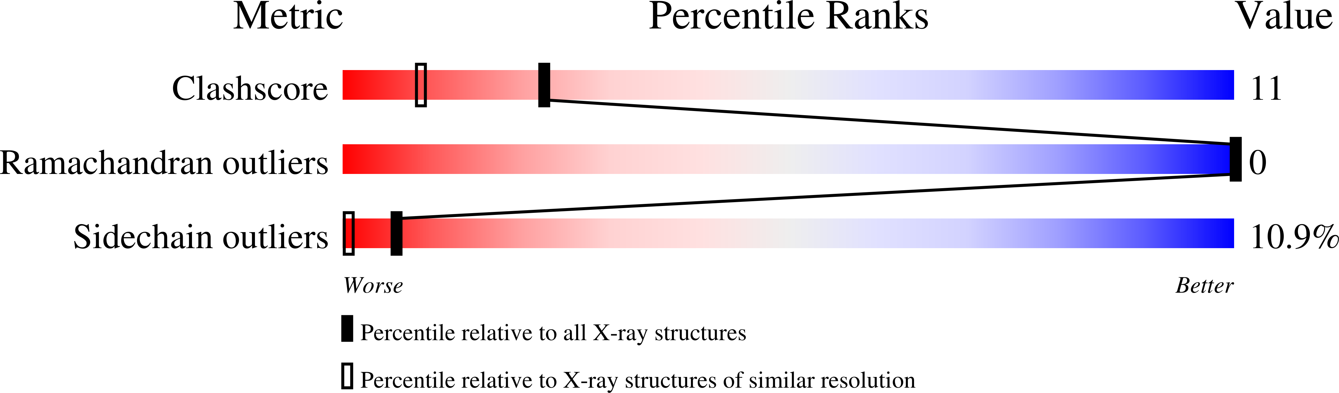
Experiments and Validation Details

Spacegroup:
P3121
Expression system: Not provided