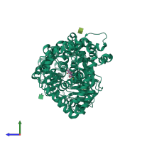
1r1j

X-ray diffraction
2.35Å resolution

STRUCTURAL ANALYSIS OF NEPRILYSIN WITH VARIOUS SPECIFIC AND POTENT INHIBITORS

Released:
Source organism: Homo sapiens
Primary publication:
Structural analysis of neprilysin with various specific and potent inhibitors.
Acta Crystallogr D Biol Crystallogr 60 392-6 (2004)
PMID: 14747736

Function and Biology Details

Reaction catalysed:
Preferential cleavage of polypeptides between hydrophobic residues, particularly with Phe or Tyr at P1'.
Biochemical function:
Cellular component:

Structure analysis Details

Assembly composition:
monomeric (preferred)
Assembly name:
PDBe Complex ID:
PDB-CPX-140129 (preferred)
Entry contents:
1 distinct polypeptide molecule
Macromolecule:
Neprilysin Chain: A
Molecule details ›
Chain: A
Length: 696 amino acids
Theoretical weight: 79.53 KDa
Source organism: Homo sapiens
Expression system: Saccharomyces cerevisiae
UniProt:
  • Canonical: P08473 (Residues: 55-750; Coverage: 93%)
Gene names: EPN, MME
Sequence domains:
Structure domains:

Ligands and Environments

3 bound ligands:
No modified residues

Experiments and Validation Details

Entry percentile scores
X-ray source: ENRAF-NONIUS FR591
Spacegroup: P3221
Unit cell:
a: 107.458Å b: 107.458Å c: 111.997Å
α: 90° β: 90° γ: 120°
R-values:
R R work R free
0.225 0.222 0.283
Expression system: Saccharomyces cerevisiae