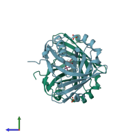
1s2p

X-ray diffraction
1.3Å resolution

The structure and refinement of apocrustacyanin C2 to 1.3A resolution and the search for differences between this protein and the homologous apoproteins A1 and C1

Released:

Function and Biology Details

Structure analysis Details

Assembly composition:
homo dimer (preferred)
PDBe Complex ID:
PDB-CPX-160331 (preferred)
Entry contents:
1 distinct polypeptide molecule
Macromolecule:
Crustacyanin-C1 subunit Chains: A, B
Molecule details ›
Chains: A, B
Length: 181 amino acids
Theoretical weight: 20.69 KDa
Source organism: Homarus gammarus
UniProt:
  • Canonical: P80029 (Residues: 1-181; Coverage: 100%)
Sequence domains: Lipocalin / cytosolic fatty-acid binding protein family
Structure domains: Lipocalin

Ligands and Environments

2 bound ligands:
No modified residues

Experiments and Validation Details

Entry percentile scores
X-ray source: SRS
Spacegroup: P212121
Unit cell:
a: 41.148Å b: 79.846Å c: 110.263Å
α: 90° β: 90° γ: 90°
R-values:
R R work R free
0.19 0.189 0.215