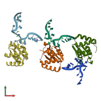
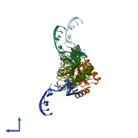
1sds

X-ray diffraction
1.8Å resolution

Structure of protein L7Ae bound to a K-turn derived from an archaeal box H/ACA sRNA

Released:

Function and Biology Details

Structure analysis Details

Assembly composition:
hetero hexamer (preferred)
PDBe Complex ID:
PDB-CPX-156939 (preferred)
Entry contents:
1 distinct polypeptide molecule
1 distinct RNA molecule
Macromolecules (2 distinct):
Large ribosomal subunit protein eL8 Chains: A, B, C
Molecule details ›
Chains: A, B, C
Length: 117 amino acids
Theoretical weight: 12.7 KDa
Source organism: Methanocaldococcus jannaschii
Expression system: Escherichia coli
UniProt:
  • Canonical: P54066 (Residues: 1-117; Coverage: 100%)
Gene names: MJ1203, rpl7ae
Sequence domains: Ribosomal protein L7Ae/L30e/S12e/Gadd45 family
Structure domains: 60s Ribosomal Protein L30; Chain: A;
box H/ACA sRNA Chains: D, E, F
Molecule details ›
Chains: D, E, F
Length: 15 nucleotides
Theoretical weight: 4.86 KDa
Source organism: Methanocaldococcus jannaschii
Expression system: Not provided

Ligands and Environments

2 bound ligands:
No modified residues

Experiments and Validation Details

Entry percentile scores
X-ray source: ALS BEAMLINE 5.0.1, ALS BEAMLINE 5.0.2
Spacegroup: F222
Unit cell:
a: 106.97Å b: 141.3Å c: 162.87Å
α: 90° β: 90° γ: 90°
R-values:
R R work R free
0.207 0.207 0.225
Expression systems:
  • Escherichia coli
  • Not provided