1t5r

X-ray diffraction
2Å resolution

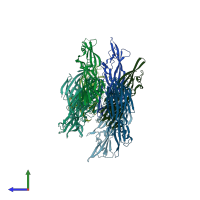
STRUCTURE OF THE PANTON-VALENTINE LEUCOCIDIN S COMPONENT FROM STAPHYLOCOCCUS AUREUS

Released:

Function and Biology Details

Biochemical function:
  • not assigned
Biological process:
Cellular component:

Structure analysis Details

Assembly composition:
homo octamer (preferred)
PDBe Complex ID:
PDB-CPX-181223 (preferred)
Entry contents:
1 distinct polypeptide molecule
Macromolecule:
Leukocidin/Hemolysin toxin domain-containing protein Chains: A, B, C, D, E, F, G, H
Molecule details ›
Chains: A, B, C, D, E, F, G, H
Length: 284 amino acids
Theoretical weight: 32.36 KDa
Source organism: Staphylococcus phage PVL
UniProt:
  • Canonical: Q783R1 (Residues: 29-312; Coverage: 91%)
Gene name: lukS-PV
Sequence domains: Leukocidin/Hemolysin toxin family
Structure domains: Leukocidin/porin MspA

Ligands and Environments

No bound ligands
No modified residues

Experiments and Validation Details

Entry percentile scores
X-ray source: ESRF BEAMLINE ID14-1
Spacegroup: P43
Unit cell:
a: 94.85Å b: 94.85Å c: 306.11Å
α: 90° β: 90° γ: 90°
R-values:
R R work R free
0.226 0.209 0.247