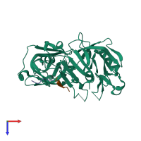
1tzs

X-ray diffraction
2.35Å resolution

Crystal Structure of an activation intermediate of Cathepsin E

Released:
Source organism: Homo sapiens

Function and Biology Details

Reaction catalysed:
Similar to cathepsin D, but slightly broader specificity.
Biological process:

Structure analysis Details

Assembly composition:
hetero hexamer (preferred)
Assembly name:
PDBe Complex ID:
PDB-CPX-146853 (preferred)
Entry contents:
3 distinct polypeptide molecules
Macromolecules (3 distinct):
Cathepsin E Chain: A
Molecule details ›
Chain: A
Length: 351 amino acids
Theoretical weight: 37.66 KDa
Source organism: Homo sapiens
Expression system: Escherichia coli
UniProt:
  • Canonical: P14091 (Residues: 54-396; Coverage: 91%)
Gene name: CTSE
Sequence domains: Eukaryotic aspartyl protease
Structure domains: Acid Proteases
Cathepsin E Chain: P
Molecule details ›
Chain: P
Length: 35 amino acids
Theoretical weight: 4.22 KDa
Source organism: Homo sapiens
Expression system: Escherichia coli
UniProt:
  • Canonical: P14091 (Residues: 19-53; Coverage: 9%)
Gene name: CTSE
Sequence domains: A1 Propeptide
23-mer peptide from PelB-IgG kappa light chain fusion protein Chain: X
Molecule details ›
Chain: X
Length: 23 amino acids
Theoretical weight: 2.36 KDa

Ligands and Environments

No bound ligands
No modified residues

Experiments and Validation Details

Entry percentile scores
X-ray source: ENRAF-NONIUS FR591
Spacegroup: P41212
Unit cell:
a: 61.321Å b: 61.321Å c: 207.8Å
α: 90° β: 90° γ: 90°
R-values:
R R work R free
0.198 0.198 0.238
Expression system: Escherichia coli