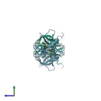
1xqh

X-ray diffraction
1.75Å resolution

Crystal structure of a ternary complex of the methyltransferase SET9 (also known as SET7/9) with a P53 peptide and SAH

Released:

Function and Biology Details

Reaction catalysed:
S-adenosyl-L-methionine + a [histone H3]-L-lysine(4) = S-adenosyl-L-homocysteine + a [histone H3]-N(6)-methyl-L-lysine(4)
Cellular component:

Structure analysis Details

Assembly composition:
hetero dimer (preferred)
PDBe Complex ID:
PDB-CPX-138121 (preferred)
Entry contents:
2 distinct polypeptide molecules
Macromolecules (2 distinct):
Histone-lysine N-methyltransferase SETD7 Chains: A, E
Molecule details ›
Chains: A, E
Length: 264 amino acids
Theoretical weight: 29.32 KDa
Source organism: Homo sapiens
Expression system: Escherichia coli BL21(DE3)
UniProt:
  • Canonical: Q8WTS6 (Residues: 108-366; Coverage: 71%)
Gene names: KIAA1717, KMT7, SET7, SET9, SETD7
Sequence domains:
Structure domains:
Cellular tumor antigen p53 Chains: B, F
Molecule details ›
Chains: B, F
Length: 10 amino acids
Theoretical weight: 1.16 KDa
Source organism: Homo sapiens
Expression system: Not provided
UniProt:
  • Canonical: P04637 (Residues: 369-377; Coverage: 2%)
Gene names: P53, TP53

Ligands and Environments

1 bound ligand:
1 modified residue:

Experiments and Validation Details

Entry percentile scores
X-ray source: SRS BEAMLINE PX14.2
Spacegroup: P21
Unit cell:
a: 40.374Å b: 103.123Å c: 67.165Å
α: 90° β: 90.04° γ: 90°
R-values:
R R work R free
0.186 0.184 0.223
Expression systems:
  • Escherichia coli BL21(DE3)
  • Not provided