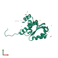
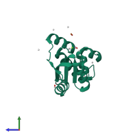
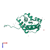
1zd0

X-ray diffraction
1.7Å resolution

Crystal structure of Pfu-542154 conserved hypothetical protein

Released:
Source organism: Pyrococcus furiosus
Entry authors: Habel JE, Liu ZJ, Horanyi PS, Florence QJT, Tempel W, Zhou W, Chen L, Lee D, Nguyen J, Chang SH, Bereton P, Izumi M, Jenny Jr FE, Poole II FL, Shah C, Sugar FJ, Adams MWW, Rose JP, Wang BC, Southeast Collaboratory for Structural Genomics (SECSG)

Function and Biology Details

Biochemical function:
Biological process:
  • not assigned
Cellular component:
  • not assigned
Structure domain:

Structure analysis Details

Assembly composition:
monomeric (preferred)
PDBe Complex ID:
PDB-CPX-186457 (preferred)
Entry contents:
1 distinct polypeptide molecule
Macromolecule:
KEOPS complex subunit Cgi121 Chain: A
Molecule details ›
Chain: A
Length: 150 amino acids
Theoretical weight: 16.85 KDa
Source organism: Pyrococcus furiosus
Expression system: Escherichia coli BL21(DE3)
UniProt:
  • Canonical: Q8U3E5 (Residues: 2-142; Coverage: 99%)
Gene name: PF0523
Sequence domains: Kinase binding protein CGI-121
Structure domains: CGI121/TPRKB

Ligands and Environments

3 bound ligands:
No modified residues

Experiments and Validation Details

Entry percentile scores
X-ray source: APS BEAMLINE 22-ID, RIGAKU MICROMAX-007, RIGAKU FR-D
Spacegroup: P3121
Unit cell:
a: 53.46Å b: 53.46Å c: 85.7Å
α: 90° β: 90° γ: 120°
R-values:
R R work R free
0.205 0.202 0.252
Expression system: Escherichia coli BL21(DE3)