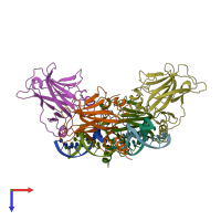
2ac0

X-ray diffraction
1.8Å resolution

Structural Basis of DNA Recognition by p53 Tetramers (complex I)

Released:
Source organism: Homo sapiens

Function and Biology Details

Structure analysis Details

Assembly composition:
hetero octamer (preferred)
PDBe Complex ID:
PDB-CPX-115065 (preferred)
Entry contents:
1 distinct polypeptide molecule
1 distinct DNA molecule
Macromolecules (2 distinct):
Cellular tumor antigen p53 Chains: A, B, C, D
Molecule details ›
Chains: A, B, C, D
Length: 200 amino acids
Theoretical weight: 22.51 KDa
Source organism: Homo sapiens
Expression system: Escherichia coli
UniProt:
  • Canonical: P04637 (Residues: 94-293; Coverage: 51%)
Gene names: P53, TP53
Sequence domains: P53 DNA-binding domain
Structure domains: Immunoglobulin-like
5'-D(*CP*GP*GP*GP*CP*AP*TP*GP*CP*CP*CP*G)-3' Chains: E, F, G, H
Molecule details ›
Chains: E, F, G, H
Length: 12 nucleotides
Theoretical weight: 3.66 KDa
Source organism: Homo sapiens
Expression system: Not provided

Ligands and Environments

1 bound ligand:
No modified residues

Experiments and Validation Details

Entry percentile scores
X-ray source: RIGAKU RUH3R
Spacegroup: P1
Unit cell:
a: 54.439Å b: 58.146Å c: 77.539Å
α: 82.93° β: 87.99° γ: 73.6°
R-values:
R R work R free
0.157 0.153 0.217
Expression systems:
  • Escherichia coli
  • Not provided