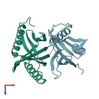
2ej8

X-ray diffraction
1.84Å resolution

Crystal structure of APPL1 PTB domain at 1.8A

Released:
Source organism: Homo sapiens
Entry authors: Tanabe H, Hosaka T, Murayama K, Terada T, Shirouzu M, Yokoyama S, RIKEN Structural Genomics/Proteomics Initiative (RSGI)

Function and Biology Details

Biochemical function:
  • not assigned
Biological process:
  • not assigned
Cellular component:
  • not assigned

Structure analysis Details

Assembly composition:
homo dimer (preferred)
PDBe Complex ID:
PDB-CPX-193847 (preferred)
Entry contents:
1 distinct polypeptide molecule
Macromolecule:
DCC-interacting protein 13-alpha Chains: A, B
Molecule details ›
Chains: A, B
Length: 160 amino acids
Theoretical weight: 18.05 KDa
Source organism: Homo sapiens
Expression system: Not provided
UniProt:
  • Canonical: Q9UKG1 (Residues: 492-644; Coverage: 22%)
Gene names: APPL, APPL1, DIP13A, KIAA1428
Sequence domains: Phosphotyrosine interaction domain (PTB/PID)
Structure domains: Pleckstrin-homology domain (PH domain)/Phosphotyrosine-binding domain (PTB)

Ligands and Environments

No bound ligands
No modified residues

Experiments and Validation Details

Entry percentile scores
X-ray source: PHOTON FACTORY BEAMLINE AR-NW12A
Spacegroup: P21
Unit cell:
a: 45.23Å b: 63.566Å c: 52.449Å
α: 90° β: 102.67° γ: 90°
R-values:
R R work R free
0.195 0.193 0.23
Expression system: Not provided