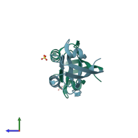
2f5y

X-ray diffraction
2.39Å resolution

Crystal Structure of the PDZ Domain from Human RGS-3

Released:
Source organism: Homo sapiens
Entry authors: Ugochukwu E, Berridge G, Johansson C, Smee C, Savitsky P, Burgess N, Colebrook S, Yang X, Elkins J, Doyle D, Turnbull A, Papagrigoriou E, Debreczeni J, Bunkoczi G, Gorrec F, von Delft F, Arrowsmith C, Sundstrom M, Weigelt J, Edwards A, Structural Genomics Consortium (SGC)

Function and Biology Details

Biochemical function:
  • not assigned
Biological process:
  • not assigned
Cellular component:
  • not assigned
Sequence domains:
Structure domain:

Structure analysis Details

Assembly composition:
monomeric (preferred)
PDBe Complex ID:
PDB-CPX-156046 (preferred)
Entry contents:
1 distinct polypeptide molecule
Macromolecule:
Regulator of G-protein signaling 3 Chains: A, B
Molecule details ›
Chains: A, B
Length: 91 amino acids
Theoretical weight: 10.24 KDa
Source organism: Homo sapiens
Expression system: Escherichia coli
UniProt:
  • Canonical: P49796 (Residues: 299-384; Coverage: 7%)
Gene name: RGS3
Sequence domains: PDZ domain
Structure domains: Pdz3 Domain

Ligands and Environments

1 bound ligand:
No modified residues

Experiments and Validation Details

Entry percentile scores
X-ray source: SLS BEAMLINE X10SA
Spacegroup: P21212
Unit cell:
a: 91.11Å b: 44.14Å c: 49.857Å
α: 90° β: 90° γ: 90°
R-values:
R R work R free
0.217 0.215 0.264
Expression system: Escherichia coli