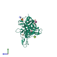
2gy5

X-ray diffraction
2.9Å resolution

Tie2 Ligand-Binding Domain Crystal Structure

Released:

Function and Biology Details

Reaction catalysed:
ATP + a [protein]-L-tyrosine = ADP + a [protein]-L-tyrosine phosphate
Biochemical function:
Cellular component:

Structure analysis Details

Assembly composition:
monomeric (preferred)
PDBe Complex ID:
PDB-CPX-169749 (preferred)
Entry contents:
1 distinct polypeptide molecule
Macromolecule:
Angiopoietin-1 receptor Chain: A
Molecule details ›
Chain: A
Length: 423 amino acids
Theoretical weight: 47.19 KDa
Source organism: Homo sapiens
Expression system: Cell free synthesis
UniProt:
  • Canonical: Q02763 (Residues: 23-445; Coverage: 38%)
Gene names: TEK, TIE2, VMCM, VMCM1
Sequence domains: Tie-2 Ig-like domain 1
Structure domains:

Ligands and Environments

3 bound ligands:
No modified residues

Experiments and Validation Details

Entry percentile scores
X-ray source: NSLS BEAMLINE X29A
Spacegroup: P41212
Unit cell:
a: 114.653Å b: 114.653Å c: 113.774Å
α: 90° β: 90° γ: 90°
R-values:
R R work R free
0.26 0.24 0.294
Expression system: Cell free synthesis