2ibf

X-ray diffraction
3.2Å resolution

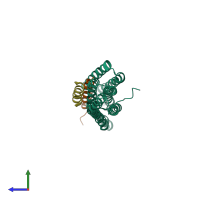
Human vinculin's head domain (Vh1, residues 1-258) in complex with two vinculin binding sites of Shigella flexneri's IpaA (residues 565-587)

Released:
Source organisms:
Primary publication:
Vinculin binding in its closed conformation by a helix addition mechanism.
EMBO J 26 4588-96 (2007)
PMID: 17932491

Function and Biology Details

Structure analysis Details

Assembly composition:
hetero trimer (preferred)
PDBe Complex ID:
PDB-CPX-148245 (preferred)
Entry contents:
2 distinct polypeptide molecules
Macromolecules (2 distinct):
Vinculin Chain: A
Molecule details ›
Chain: A
Length: 266 amino acids
Theoretical weight: 30.03 KDa
Source organism: Homo sapiens
Expression system: Escherichia coli
UniProt:
  • Canonical: P18206 (Residues: 1-258; Coverage: 23%)
Gene name: VCL
Sequence domains: Vinculin family
Structure domains: Alpha-catenin/vinculin-like
Invasin IpaA Chains: B, D
Molecule details ›
Chains: B, D
Length: 25 amino acids
Theoretical weight: 2.75 KDa
Source organism: Shigella flexneri
Expression system: Escherichia coli
UniProt:
  • Canonical: P18010 (Residues: 563-587; Coverage: 4%)
Gene names: CP0125, ipaA
Sequence domains: SipA vinculin binding site

Ligands and Environments

No bound ligands
No modified residues

Experiments and Validation Details

Entry percentile scores
X-ray source: APS BEAMLINE 19-ID
Spacegroup: P4332
Unit cell:
a: 154.478Å b: 154.478Å c: 154.478Å
α: 90° β: 90° γ: 90°
R-values:
R R work R free
0.245 0.243 0.27
Expression system: Escherichia coli