2ibg

X-ray diffraction
2.2Å resolution

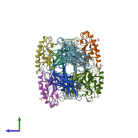
Crystal Structure of Hedgehog Bound to the FNIII Domains of Ihog

Released:
Source organism: Drosophila melanogaster
Primary publication:
Structure of a heparin-dependent complex of Hedgehog and Ihog.
Proc Natl Acad Sci U S A 103 17208-13 (2006)
PMID: 17077139

Function and Biology Details

Structure analysis Details

Assembly composition:
hetero dimer (preferred)
PDBe Complex ID:
PDB-CPX-169785 (preferred)
Entry contents:
2 distinct polypeptide molecules
Macromolecules (2 distinct):
Interference hedgehog Chains: A, B, C, D
Molecule details ›
Chains: A, B, C, D
Length: 214 amino acids
Theoretical weight: 24.29 KDa
Source organism: Drosophila melanogaster
Expression system: Escherichia coli
UniProt:
  • Canonical: Q9VM64 (Residues: 466-677; Coverage: 25%)
Gene names: CG9211, ihog
Sequence domains: Fibronectin type III domain
Structure domains: Immunoglobulins
Protein hedgehog N-product Chains: E, F, G, H
Molecule details ›
Chains: E, F, G, H
Length: 150 amino acids
Theoretical weight: 17.27 KDa
Source organism: Drosophila melanogaster
Expression system: Escherichia coli
UniProt:
  • Canonical: Q02936 (Residues: 99-248; Coverage: 32%)
Gene names: CG4637, hh
Sequence domains: Hedgehog amino-terminal signalling domain
Structure domains: Muramoyl-pentapeptide Carboxypeptidase; domain 2

Ligands and Environments

1 bound ligand:
No modified residues

Experiments and Validation Details

Entry percentile scores
X-ray source: NSLS BEAMLINE X4A
Spacegroup: P21
Unit cell:
a: 75.354Å b: 70.003Å c: 155.695Å
α: 90° β: 90.18° γ: 90°
R-values:
R R work R free
0.2 0.198 0.246
Expression system: Escherichia coli