2lw9

Solution NMR

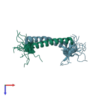
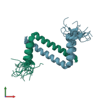
NMR solution structure of Myo10 anti-CC

Released:
Source organism: Homo sapiens
Primary publication:
Antiparallel coiled-coil-mediated dimerization of myosin X.
Proc Natl Acad Sci U S A 109 17388-93 (2012)
PMID: 23012428

Function and Biology Details

Biochemical function:
  • not assigned
Biological process:
  • not assigned
Cellular component:
  • not assigned

Structure analysis Details

Assembly composition:
homo dimer (preferred)
PDBe Complex ID:
PDB-CPX-190808 (preferred)
Entry contents:
1 distinct polypeptide molecule
Macromolecule:
Unconventional myosin-X Chains: A, B
Molecule details ›
Chains: A, B
Length: 51 amino acids
Theoretical weight: 6.35 KDa
Source organism: Homo sapiens
Expression system: Escherichia coli
UniProt:
  • Canonical: Q9HD67 (Residues: 883-933; Coverage: 3%)
Gene names: KIAA0799, MYO10
Sequence domains: Unconventional myosin-X coiled coil domain
Structure domains: Single alpha-helices involved in coiled-coils or other helix-helix interfaces

Ligands and Environments

No bound ligands
No modified residues

Experiments and Validation Details

Entry percentile scores
Chemical shift assignment: 39%
Refinement method: DGSA-distance geometry simulated annealing
Expression system: Escherichia coli