2p32

X-ray diffraction
3.2Å resolution

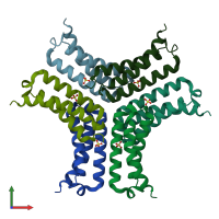
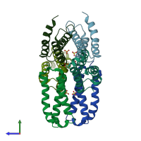
Crystal structure of the C-terminal 10 kDa subdomain from C. elegans Hsp70

Released:
Source organism: Caenorhabditis elegans
Primary publication:
Crystal structure of the C-terminal three-helix bundle subdomain of C. elegans Hsp70.
Biochem Biophys Res Commun 357 105-10 (2007)
PMID: 17407764

Function and Biology Details

Biochemical function:
  • not assigned
Biological process:
  • not assigned
Cellular component:
  • not assigned

Structure analysis Details

Assemblies composition:
monomeric (preferred)
homo hexamer
PDBe Complex ID:
PDB-CPX-140655 (preferred)
Entry contents:
1 distinct polypeptide molecule
Macromolecule:
Heat shock protein hsp-1 Chains: A, B, C, D, E, F
Molecule details ›
Chains: A, B, C, D, E, F
Length: 120 amino acids
Theoretical weight: 12.96 KDa
Source organism: Caenorhabditis elegans
Expression system: Escherichia coli
UniProt:
  • Canonical: P09446 (Residues: 542-640; Coverage: 16%)
Gene names: F26D10.3, hsp-1, hsp70a
Structure domains: Substrate Binding Domain Of Dnak; Chain:A; Domain 2

Ligands and Environments

1 bound ligand:
No modified residues

Experiments and Validation Details

Entry percentile scores
X-ray source: ESRF BEAMLINE BM14
Spacegroup: P42212
Unit cell:
a: 138.927Å b: 138.927Å c: 100.704Å
α: 90° β: 90° γ: 90°
R-values:
R R work R free
0.269 0.268 0.282
Expression system: Escherichia coli