Function and Biology Details
Reaction catalysed:
Hydrolysis of proteins with broad specificity for peptide bonds. Preferentially cleaves -Arg-Arg-|- bonds in small molecule substrates (thus differing from cathepsin L). In addition to being an endopeptidase, shows peptidyl-dipeptidase activity, liberating C-terminal dipeptides.
Biochemical function:
Biological process:
Cellular component:
Structure analysis Details
Assembly composition:
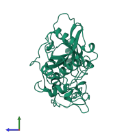
monomeric (preferred)
Assembly name:
Cathepsin B heavy chain (preferred)
PDBe Complex ID:
PDB-CPX-139795 (preferred)
Entry contents:
1 distinct polypeptide molecule
Macromolecule:
Ligands and Environments
No bound ligands
No modified residues
Experiments and Validation Details
X-ray source:
RIGAKU RUH2R
Spacegroup:
P6522
Expression system: Escherichia coli


