2v5f

X-ray diffraction
2.03Å resolution

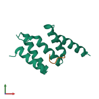
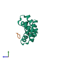
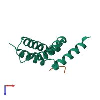
Crystal structure of wild type peptide-binding domain of human type I collagen prolyl 4-hydroxylase.

Released:
Entry authors: Pekkala M, Hieta R, Kivirikko K, Myllyharju J, Wierenga R

Function and Biology Details

Reaction catalysed:
L-proline-[procollagen] + 2-oxoglutarate + O(2) = trans-4-hydroxy-L-proline-[procollagen] + succinate + CO(2)
Biochemical function:
  • not assigned
Biological process:
  • not assigned
Cellular component:
  • not assigned

Structure analysis Details

Assembly composition:
hetero dimer (preferred)
PDBe Complex ID:
PDB-CPX-146692 (preferred)
Entry contents:
2 distinct polypeptide molecules
Macromolecules (2 distinct):
Prolyl 4-hydroxylase subunit alpha-1 Chain: A
Molecule details ›
Chain: A
Length: 104 amino acids
Theoretical weight: 12.08 KDa
Source organism: Homo sapiens
Expression system: Escherichia coli BL21(DE3)
UniProt:
  • Canonical: P13674 (Residues: 161-263; Coverage: 20%)
Gene names: P4HA, P4HA1
Structure domains: Tetratricopeptide repeat domain
HEXA-HISTIDINE PEPTIDE Chain: X
Molecule details ›
Chain: X
Length: 6 amino acids
Theoretical weight: 847 Da
Source organism: synthetic construct
Expression system: Escherichia coli BL21(DE3)

Ligands and Environments

No bound ligands
No modified residues

Experiments and Validation Details

Entry percentile scores
X-ray source: ENRAF-NONIUS FR591
Spacegroup: P212121
Unit cell:
a: 37.472Å b: 59.046Å c: 61.668Å
α: 90° β: 90° γ: 90°
R-values:
R R work R free
0.199 0.196 0.265
Expression system: Escherichia coli BL21(DE3)