2zmv

X-ray diffraction
2.8Å resolution

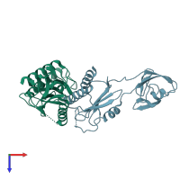
Crystal structure of Synbindin

Released:
Source organism: Homo sapiens
Primary publication:
Crystal structure of human synbindin reveals two conformations of longin domain.
Biochem Biophys Res Commun 378 338-43 (2009)
PMID: 18466758

Function and Biology Details

Structure analysis Details

Assemblies composition:
homo hexamer
homo dimer (preferred)
PDBe Complex ID:
PDB-CPX-195120 (preferred)
Entry contents:
1 distinct polypeptide molecule
Macromolecule:
Trafficking protein particle complex subunit 4 Chains: A, B
Molecule details ›
Chains: A, B
Length: 227 amino acids
Theoretical weight: 25.44 KDa
Source organism: Homo sapiens
Expression system: Escherichia coli
UniProt:
  • Canonical: Q9Y296 (Residues: 1-219; Coverage: 100%)
Gene names: CGI-104, HSPC172, PTD009, SBDN, TRAPPC4
Sequence domains: Sybindin-like family
Structure domains: Beta-Lactamase

Ligands and Environments

No bound ligands
No modified residues

Experiments and Validation Details

Entry percentile scores
X-ray source: BSRF BEAMLINE 3W1A, RIGAKU FR-E+ DW
Spacegroup: P213
Unit cell:
a: 124.391Å b: 124.391Å c: 124.391Å
α: 90° β: 90° γ: 90°
R-values:
R R work R free
0.245 0.243 0.29
Expression system: Escherichia coli