2gs0

Solution NMR

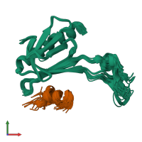
NMR structure of the complex between the PH domain of the Tfb1 subunit from TFIIH and the activation domain of p53

Released:

Function and Biology Details

Biochemical function:
  • not assigned
Biological process:
  • not assigned
Cellular component:
  • not assigned

Structure analysis Details

Assembly composition:
hetero dimer (preferred)
PDBe Complex ID:
PDB-CPX-138109 (preferred)
Entry contents:
2 distinct polypeptide molecules
Macromolecules (2 distinct):
General transcription and DNA repair factor IIH subunit TFB1 Chain: A
Molecule details ›
Chain: A
Length: 115 amino acids
Theoretical weight: 12.9 KDa
Source organism: Saccharomyces cerevisiae
Expression system: Escherichia coli
UniProt:
  • Canonical: P32776 (Residues: 1-115; Coverage: 18%)
Gene names: D9740.3, TFB1, YDR311W
Sequence domains: TFIIH p62 subunit, N-terminal domain
Structure domains: Pleckstrin-homology domain (PH domain)/Phosphotyrosine-binding domain (PTB)
Cellular tumor antigen p53 Chain: B
Molecule details ›
Chain: B
Length: 54 amino acids
Theoretical weight: 6.02 KDa
Source organism: Homo sapiens
Expression system: Escherichia coli
UniProt:
  • Canonical: P04637 (Residues: 20-73; Coverage: 14%)
Gene names: P53, TP53
Sequence domains:

Ligands and Environments

No bound ligands
No modified residues

Experiments and Validation Details

Entry percentile scores
Refinement method: Simulated annealing, with a combination of torsion angle and Cartesian dynamics.
Expression system: Escherichia coli