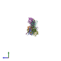
2hod

X-ray diffraction
2.9Å resolution

Crystal Structure of Fragment D from Human Fibrinogen Complexed with Gly-hydroxyPro-Arg-Pro-amide

Released:
Source organism: Homo sapiens
Entry authors: Doolittle RF, Kollman JM, Chen A, Pandi L

Function and Biology Details

Structure analysis Details

Assembly composition:
hetero pentamer (preferred)
PDBe Complex ID:
PDB-CPX-136008 (preferred)
Entry contents:
4 distinct polypeptide molecules
Macromolecules (4 distinct):
Fibrinogen alpha chain Chains: A, D, G, J
Molecule details ›
Chains: A, D, G, J
Length: 87 amino acids
Theoretical weight: 10.24 KDa
Source organism: Homo sapiens
UniProt:
  • Canonical: P02671 (Residues: 130-216; Coverage: 10%)
Gene name: FGA
Sequence domains: Fibrinogen alpha/beta chain family
Structure domains: Single alpha-helices involved in coiled-coils or other helix-helix interfaces
Fibrinogen beta chain Chains: B, E, H, K
Molecule details ›
Chains: B, E, H, K
Length: 328 amino acids
Theoretical weight: 37.69 KDa
Source organism: Homo sapiens
UniProt:
  • Canonical: P02675 (Residues: 164-491; Coverage: 71%)
Gene name: FGB
Sequence domains:
Structure domains:
Fibrinogen gamma chain Chains: C, F, I, L
Molecule details ›
Chains: C, F, I, L
Length: 323 amino acids
Theoretical weight: 36.56 KDa
Source organism: Homo sapiens
UniProt:
  • Canonical: P02679 (Residues: 115-433; Coverage: 75%)
Gene names: FGG, PRO2061
Sequence domains:
Structure domains:
Gly-hydroxyPro-Arg-Pro-amide peptide ligand Chains: M, N, O, P, Q, R, S, T
Molecule details ›
Chains: M, N, O, P, Q, R, S, T
Length: 5 amino acids
Theoretical weight: 440 Da

Ligands and Environments

2 bound ligands:
1 modified residue:

Experiments and Validation Details

Entry percentile scores
X-ray source: ALS BEAMLINE 8.2.2
Spacegroup: P2
Unit cell:
a: 81.647Å b: 47.069Å c: 431.46Å
α: 90° β: 90.06° γ: 90°
R-values:
R R work R free
0.244 0.268 0.347