


Function and Biology Details
Biochemical function:
- not assigned
Biological process:
Sequence domains:
Structure domain:
Structure analysis Details
Assembly composition:
monomeric (preferred)
Assembly name:
Leu/Ile/Val-binding protein (preferred)
PDBe Complex ID:
PDB-CPX-142337 (preferred)
Entry contents:
1 distinct polypeptide molecule
Macromolecule:
Leu/Ile/Val-binding protein
Molecule details ›
Chain: A
Length: 344 amino acids
Theoretical weight: 36.78 KDa
Source organism: Escherichia coli
Expression system: Not provided
UniProt:
Sequence domains: Periplasmic binding protein
Structure domains: Rossmann fold
Length: 344 amino acids
Theoretical weight: 36.78 KDa
Source organism: Escherichia coli
Expression system: Not provided
UniProt:
- Canonical:
 P0AD96 (Residues: 24-367; Coverage: 100%)
Sequence domains: Periplasmic binding protein
Structure domains: Rossmann fold
Ligands and Environments
No bound ligands
No modified residues
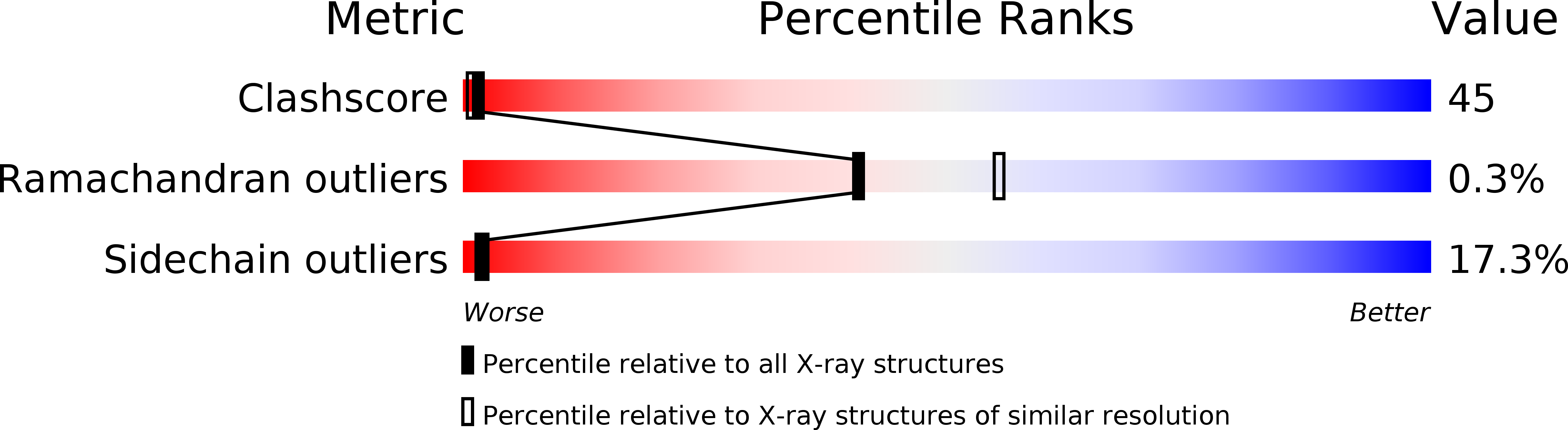
Experiments and Validation Details

Spacegroup:
P212121
Expression system: Not provided