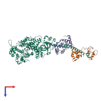
2os8

X-ray diffraction
3.27Å resolution

Rigor-like structures of muscle myosins reveal key mechanical elements in the transduction pathways of this allosteric motor

Released:

Function and Biology Details

Structure analysis Details

Assembly composition:
hetero trimer (preferred)
PDBe Complex ID:
PDB-CPX-173237 (preferred)
Entry contents:
3 distinct polypeptide molecules
Macromolecules (3 distinct):
Myosin heavy chain Chain: A
Molecule details ›
Chain: A
Length: 840 amino acids
Theoretical weight: 96.14 KDa
Source organism: Placopecten magellanicus
UniProt:
  • Canonical: Q26080 (Residues: 1-840; Coverage: 43%)
Sequence domains:
EF-hand domain-containing protein Chain: B
Molecule details ›
Chain: B
Length: 157 amino acids
Theoretical weight: 17.77 KDa
Source organism: Placopecten magellanicus
UniProt:
  • Canonical: Q26068 (Residues: 1-157; Coverage: 100%)
Sequence domains: EF-hand domain pair
Structure domains: EF-hand
EF-hand domain-containing protein Chain: C
Molecule details ›
Chain: C
Length: 157 amino acids
Theoretical weight: 17.81 KDa
Source organism: Placopecten magellanicus
UniProt:
  • Canonical: Q26066 (Residues: 1-157; Coverage: 100%)
Sequence domains: EF-hand domain pair
Structure domains: EF-hand

Ligands and Environments

2 bound ligands:
No modified residues

Experiments and Validation Details

Entry percentile scores
X-ray source: CHESS BEAMLINE A1
Spacegroup: P21
Unit cell:
a: 85.567Å b: 50.359Å c: 161.621Å
α: 90° β: 98.72° γ: 90°
R-values:
R R work R free
0.281 0.281 0.314