2p5t

X-ray diffraction
3.2Å resolution

Molecular and structural characterization of the PezAT chromosomal toxin-antitoxin system of the human pathogen Streptococcus pneumoniae

Released:

Function and Biology Details

Reaction catalysed:
ATP + UDP-N-acetyl-alpha-D-glucosamine = ADP + UDP-N-acetyl-alpha-D-glucosamine 3'-phosphate
Biochemical function:
Biological process:
  • not assigned
Cellular component:
  • not assigned

Structure analysis Details

Assemblies composition:
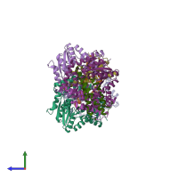
hetero tetramer
hetero pentamer (preferred)
PDBe Complex ID:
PDB-CPX-163143 (preferred)
Entry contents:
3 distinct polypeptide molecules
Macromolecules (3 distinct):
fragment of PezA helix-turn-helix motif Chain: X
Molecule details ›
Chain: X
Length: 33 amino acids
Theoretical weight: 2.83 KDa
Source organism: Streptococcus pneumoniae
Expression system: Escherichia coli
Antitoxin PezA Chains: A, C, E, G
Molecule details ›
Chains: A, C, E, G
Length: 158 amino acids
Theoretical weight: 18.27 KDa
Source organism: Streptococcus pneumoniae TIGR4
Expression system: Escherichia coli
UniProt:
  • Canonical: Q97QZ2 (Residues: 1-158; Coverage: 100%)
Gene names: SP_1050, pezA
Sequence domains: Helix-turn-helix
Structure domains: Helicase, Ruva Protein; domain 3
Toxin PezT Chains: B, D, F, H
Molecule details ›
Chains: B, D, F, H
Length: 253 amino acids
Theoretical weight: 29.11 KDa
Source organism: Streptococcus pneumoniae
Expression system: Escherichia coli
UniProt:
  • Canonical: Q97QZ1 (Residues: 1-253; Coverage: 100%)
Gene names: SP_1051, pezT
Sequence domains: Zeta toxin
Structure domains: P-loop containing nucleotide triphosphate hydrolases

Ligands and Environments

No bound ligands
No modified residues

Experiments and Validation Details

Entry percentile scores
X-ray source: SLS BEAMLINE X10SA
Spacegroup: P212121
Unit cell:
a: 80.52Å b: 102.86Å c: 254.44Å
α: 90° β: 90° γ: 90°
R-values:
R R work R free
0.217 0.214 0.277
Expression system: Escherichia coli