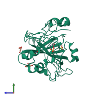
2vvb

X-ray diffraction
1.66Å resolution

Human carbonic anhydrase II in complex with bicarbonate

Released:
Source organism: Homo sapiens
Primary publication:
Structural study of X-ray induced activation of carbonic anhydrase.
Proc Natl Acad Sci U S A 106 10609-13 (2009)
PMID: 19520834

Function and Biology Details

Reactions catalysed:
H(2)CO(3) = CO(2) + H(2)O
Urea = cyanamide + H(2)O
Biochemical function:
Cellular component:

Structure analysis Details

Assembly composition:
monomeric (preferred)
Assembly name:
PDBe Complex ID:
PDB-CPX-133681 (preferred)
Entry contents:
1 distinct polypeptide molecule
Macromolecule:
Carbonic anhydrase 2 Chain: X
Molecule details ›
Chain: X
Length: 260 amino acids
Theoretical weight: 29.29 KDa
Source organism: Homo sapiens
Expression system: Escherichia coli
UniProt:
  • Canonical: P00918 (Residues: 1-260; Coverage: 100%)
Gene name: CA2
Sequence domains: Eukaryotic-type carbonic anhydrase
Structure domains: Alpha carbonic anhydrase

Ligands and Environments

No modified residues

Experiments and Validation Details

Entry percentile scores
X-ray source: ELETTRA BEAMLINE 5.2R
Spacegroup: P21
Unit cell:
a: 42.667Å b: 41.653Å c: 72.91Å
α: 90° β: 104.79° γ: 90°
R-values:
R R work R free
0.171 0.168 0.213
Expression system: Escherichia coli