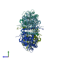
2xsn

X-ray diffraction
2.68Å resolution

Crystal Structure of Human Tyrosine Hydroxylase Catalytic Domain

Released:
Source organism: Homo sapiens
Entry authors: Muniz JRC, Cooper CDO, Yue WW, Krysztofinska E, von Delft F, Knapp S, Gileadi O, Arrowsmith CH, Edwards AM, Weigelt J, Bountra C, Kavanagh KL, Oppermann U

Function and Biology Details

Structure analysis Details

Assembly composition:
homo tetramer (preferred)
PDBe Complex ID:
PDB-CPX-139372 (preferred)
Entry contents:
1 distinct polypeptide molecule
Macromolecule:
Tyrosine 3-monooxygenase Chains: A, B, C, D
Molecule details ›
Chains: A, B, C, D
Length: 343 amino acids
Theoretical weight: 39.08 KDa
Source organism: Homo sapiens
Expression system: Escherichia coli BL21(DE3)
UniProt:
  • Canonical: P07101 (Residues: 193-528; Coverage: 64%)
Gene names: TH, TYH
Sequence domains: Biopterin-dependent aromatic amino acid hydroxylase
Structure domains: Aromatic amino acid hydroxylase

Ligands and Environments

1 bound ligand:
No modified residues

Experiments and Validation Details

Entry percentile scores
X-ray source: DIAMOND BEAMLINE I02
Spacegroup: P6522
Unit cell:
a: 191.42Å b: 191.42Å c: 168.16Å
α: 90° β: 90° γ: 120°
R-values:
R R work R free
0.191 0.189 0.222
Expression system: Escherichia coli BL21(DE3)