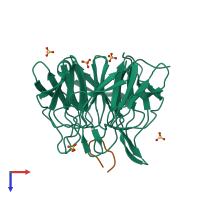
2z32

X-ray diffraction
2Å resolution

Crystal structure of Keap1 complexed with Prothymosin alpha

Released:
Source organism: Mus musculus
Primary publication:
Structural analysis of the complex of Keap1 with a prothymosin alpha peptide.
Acta Crystallogr Sect F Struct Biol Cryst Commun 64 233-8 (2008)
PMID: 18391415

Function and Biology Details

Biochemical function:
  • not assigned
Biological process:
  • not assigned
Cellular component:
  • not assigned
Structure domain:

Structure analysis Details

Assembly composition:
hetero dimer (preferred)
PDBe Complex ID:
PDB-CPX-150710 (preferred)
Entry contents:
2 distinct polypeptide molecules
Macromolecules (2 distinct):
Kelch-like ECH-associated protein 1 Chain: A
Molecule details ›
Chain: A
Length: 318 amino acids
Theoretical weight: 35.01 KDa
Source organism: Mus musculus
Expression system: Escherichia coli
UniProt:
  • Canonical: Q9Z2X8 (Residues: 308-624; Coverage: 51%)
Gene names: Inrf2, Keap1, Kiaa0132
Sequence domains: Kelch motif
Structure domains: Kelch-type beta propeller
Prothymosin alpha, N-terminally processed Chain: B
Molecule details ›
Chain: B
Length: 16 amino acids
Theoretical weight: 1.79 KDa
Source organism: Mus musculus
Expression system: Not provided
UniProt:
  • Canonical: P26350 (Residues: 39-54; Coverage: 14%)
Gene name: Ptma

Ligands and Environments

1 bound ligand:
No modified residues

Experiments and Validation Details

Entry percentile scores
X-ray source: PHOTON FACTORY BEAMLINE BL-5A
Spacegroup: P61
Unit cell:
a: 103.515Å b: 103.515Å c: 56.16Å
α: 90° β: 90° γ: 120°
R-values:
R R work R free
0.169 0.166 0.223
Expression systems:
  • Escherichia coli
  • Not provided