2zps

X-ray diffraction
1.55Å resolution

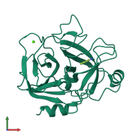
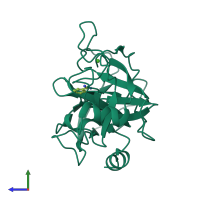
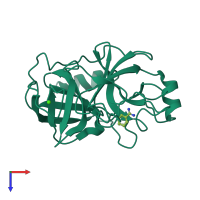
Crystal structure of anionic trypsin isoform 3 from chum salmon

Released:
Source organism: Oncorhynchus keta
Primary publication:
A structural comparison of three isoforms of anionic trypsin from chum salmon (Oncorhynchus keta).
Acta Crystallogr D Biol Crystallogr 65 717-23 (2009)
PMID: 19564692

Function and Biology Details

Structure analysis Details

Assembly composition:
monomeric (preferred)
PDBe Complex ID:
PDB-CPX-109979 (preferred)
Entry contents:
1 distinct polypeptide molecule
Macromolecule:
Peptidase S1 domain-containing protein Chain: A
Molecule details ›
Chain: A
Length: 222 amino acids
Theoretical weight: 23.89 KDa
Source organism: Oncorhynchus keta
UniProt:
  • Canonical: B3Y8K6 (Residues: 1-222; Coverage: 100%)
Sequence domains: Trypsin
Structure domains: Trypsin-like serine proteases

Ligands and Environments

2 bound ligands:
No modified residues

Experiments and Validation Details

Entry percentile scores
X-ray source: RIGAKU MICROMAX-007
Spacegroup: P212121
Unit cell:
a: 44.371Å b: 47.531Å c: 81.508Å
α: 90° β: 90° γ: 90°
R-values:
R R work R free
0.186 0.186 0.196