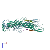
3agz

X-ray diffraction
2.51Å resolution

Crystal structure of human Hsp40 Hdj1 peptide-binding domain complexed with a C-terminal peptide of Hsp70

Released:

Function and Biology Details

Reaction catalysed:
ATP + H(2)O = ADP + phosphate
Biochemical function:
Biological process:
Cellular component:
  • not assigned

Structure analysis Details

Assembly composition:
hetero hexamer (preferred)
PDBe Complex ID:
PDB-CPX-145682 (preferred)
Entry contents:
2 distinct polypeptide molecules
Macromolecules (2 distinct):
DnaJ homolog subfamily B member 1 Chains: A, B
Molecule details ›
Chains: A, B
Length: 190 amino acids
Theoretical weight: 21.48 KDa
Source organism: Homo sapiens
Expression system: Escherichia coli
UniProt:
  • Canonical: P25685 (Residues: 151-340; Coverage: 56%)
Gene names: DNAJ1, DNAJB1, HDJ1, HSPF1
Sequence domains: DnaJ C terminal domain
Structure domains: Urease metallochaperone UreE, N-terminal domain
Heat shock cognate 71 kDa protein Chains: C, D, E, F
Molecule details ›
Chains: C, D, E, F
Length: 8 amino acids
Theoretical weight: 859 Da
Source organism: Homo sapiens
Expression system: Not provided
UniProt:
  • Canonical: P11142 (Residues: 639-646; Coverage: 1%)
Gene names: HSC70, HSP73, HSPA10, HSPA8

Ligands and Environments

No bound ligands
No modified residues

Experiments and Validation Details

Entry percentile scores
X-ray source: BRUKER AXS MICROSTAR
Spacegroup: C2
Unit cell:
a: 111.057Å b: 50.904Å c: 90.148Å
α: 90° β: 114.76° γ: 90°
R-values:
R R work R free
0.222 0.22 0.269
Expression systems:
  • Escherichia coli
  • Not provided