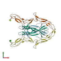
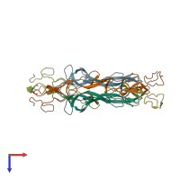
3buk

X-ray diffraction
2.6Å resolution

Crystal Structure of the Neurotrophin-3 and p75NTR Symmetrical Complex

Released:
Source organisms:
Primary publication:
Crystal structure of the neurotrophin-3 and p75NTR symmetrical complex.
Nature 454 789-93 (2008)
PMID: 18596692

Function and Biology Details

Structure analysis Details

Assembly composition:
hetero tetramer (preferred)
PDBe Complex ID:
PDB-CPX-139423 (preferred)
Entry contents:
2 distinct polypeptide molecules
Macromolecules (2 distinct):
Neurotrophin-3 Chains: A, B
Molecule details ›
Chains: A, B
Length: 119 amino acids
Theoretical weight: 13.65 KDa
Source organism: Homo sapiens
Expression system: Escherichia coli
UniProt:
  • Canonical: P20783 (Residues: 139-257; Coverage: 50%)
Gene name: NTF3
Sequence domains: Nerve growth factor family
Structure domains: Cystine-knot cytokines
Tumor necrosis factor receptor superfamily member 16 Chains: C, D
Molecule details ›
Chains: C, D
Length: 167 amino acids
Theoretical weight: 18.06 KDa
Source organism: Rattus norvegicus
Expression system: Spodoptera frugiperda
UniProt:
  • Canonical: P07174 (Residues: 30-190; Coverage: 41%)
Gene names: Ngfr, Tnfrsf16
Sequence domains: TNFR/NGFR cysteine-rich region
Structure domains: Tumor Necrosis Factor Receptor, subunit A, domain 2

Ligands and Environments

1 bound ligand:
No modified residues

Experiments and Validation Details

Entry percentile scores
X-ray source: EMBL/DESY, HAMBURG BEAMLINE BW7A
Spacegroup: R3
Unit cell:
a: 125.8Å b: 125.8Å c: 133.1Å
α: 90° β: 90° γ: 120°
R-values:
R R work R free
0.227 0.224 0.288
Expression systems:
  • Escherichia coli
  • Spodoptera frugiperda