3d3w

X-ray diffraction
1.87Å resolution

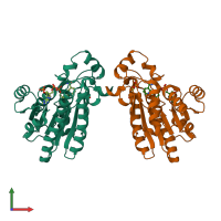
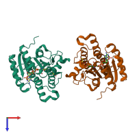
Structure of L-Xylulose Reductase with bound coenzyme, phosphate and hydroxide.

Released:

Function and Biology Details

Structure analysis Details

Assemblies composition:
hetero tetramer
hetero dimer (preferred)
homo dimer
Assembly name:
PDBe Complex ID:
PDB-CPX-182273 (preferred)
Entry contents:
2 distinct polypeptide molecules
Macromolecules (2 distinct):
L-xylulose reductase Chain: A
Molecule details ›
Chain: A
Length: 244 amino acids
Theoretical weight: 25.96 KDa
Source organism: Homo sapiens
Expression system: Escherichia coli
UniProt:
  • Canonical: Q7Z4W1 (Residues: 1-244; Coverage: 100%)
Gene names: DCXR, SDR20C1
Sequence domains: Enoyl-(Acyl carrier protein) reductase
Structure domains: NAD(P)-binding Rossmann-like Domain
L-xylulose reductase Chain: B
Molecule details ›
Chain: B
Length: 244 amino acids
Theoretical weight: 25.94 KDa
Source organism: Homo sapiens
Expression system: Escherichia coli
UniProt:
  • Canonical: Q7Z4W1 (Residues: 1-244; Coverage: 100%)
Gene names: DCXR, SDR20C1
Sequence domains: Enoyl-(Acyl carrier protein) reductase
Structure domains: NAD(P)-binding Rossmann-like Domain

Ligands and Environments


Cofactor: Ligand NAP 2 x NAP
1 bound ligand:
1 modified residue:

Experiments and Validation Details

Entry percentile scores
X-ray source: RIGAKU RU300
Spacegroup: P21212
Unit cell:
a: 73.678Å b: 87.476Å c: 72.168Å
α: 90° β: 90° γ: 90°
R-values:
R R work R free
0.207 0.204 0.258
Expression system: Escherichia coli