3dy0

X-ray diffraction
1.55Å resolution

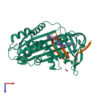
Crystal Structure of Cleaved PCI Bound to Heparin

Released:
Source organism: Homo sapiens
Primary publication:
The heparin binding site of protein C inhibitor is protease-dependent.
J Biol Chem 283 36039-45 (2008)
PMID: 18974053

Function and Biology Details

Structure analysis Details

Assembly composition:
hetero dimer (preferred)
PDBe Complex ID:
PDB-CPX-138559 (preferred)
Entry contents:
2 distinct polypeptide molecules
Macromolecules (3 distinct):
Plasma serine protease inhibitor Chain: A
Molecule details ›
Chain: A
Length: 336 amino acids
Theoretical weight: 37.48 KDa
Source organism: Homo sapiens
Expression system: Escherichia coli
UniProt:
  • Canonical: P05154 (Residues: 37-372; Coverage: 87%)
Gene names: PCI, PLANH3, PROCI, SERPINA5
Sequence domains: Serpin (serine protease inhibitor)
Structure domains:
Plasma serine protease inhibitor Chain: B
Molecule details ›
Chain: B
Length: 29 amino acids
Theoretical weight: 3.51 KDa
Source organism: Homo sapiens
Expression system: Escherichia coli
UniProt:
  • Canonical: P05154 (Residues: 379-406; Coverage: 7%)
Gene names: PCI, PLANH3, PROCI, SERPINA5

Ligands and Environments

Carbohydrate polymer : NEW Components: IDS, SGN
2 bound ligands:
No modified residues

Experiments and Validation Details

Entry percentile scores
X-ray source: SRS BEAMLINE PX14.1
Spacegroup: P21
Unit cell:
a: 61.051Å b: 49.451Å c: 64.969Å
α: 90° β: 113.08° γ: 90°
R-values:
R R work R free
0.197 0.197 0.217
Expression system: Escherichia coli