


Function and Biology Details
Biochemical function:
- not assigned
Biological process:
- not assigned
Cellular component:
- not assigned
Sequence domains:
Structure analysis Details
Assembly composition:
monomeric (preferred)
Assembly name:
Spectrin beta chain, erythrocytic (preferred)
PDBe Complex ID:
PDB-CPX-145745 (preferred)
Entry contents:
1 distinct polypeptide molecule
Macromolecule:
Spectrin beta chain, erythrocytic
Molecule details ›
Chain: A
Length: 218 amino acids
Theoretical weight: 24.81 KDa
Source organism: Homo sapiens
Expression system: Escherichia coli
UniProt:
Sequence domains: Spectrin repeat
Structure domains: Methane Monooxygenase Hydroxylase; Chain G, domain 1
Length: 218 amino acids
Theoretical weight: 24.81 KDa
Source organism: Homo sapiens
Expression system: Escherichia coli
UniProt:
- Canonical:
 P11277 (Residues: 1692-1907; Coverage: 10%)
Sequence domains: Spectrin repeat
Structure domains: Methane Monooxygenase Hydroxylase; Chain G, domain 1
Ligands and Environments
No bound ligands
No modified residues
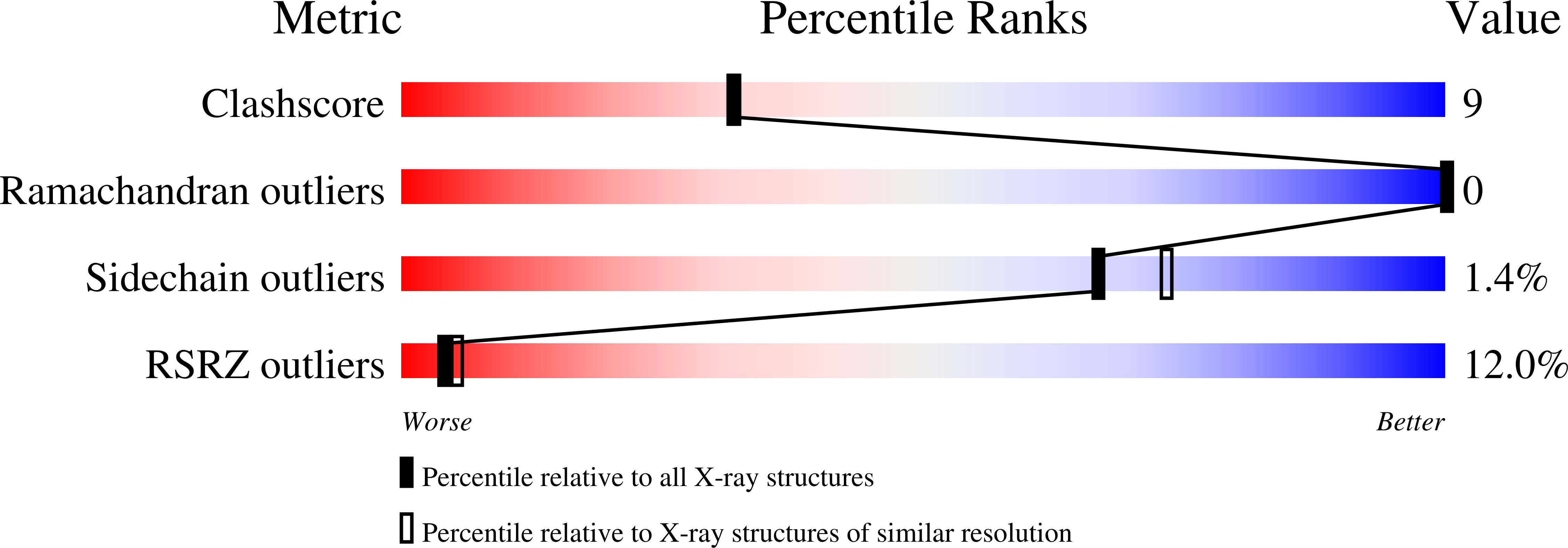
Experiments and Validation Details

X-ray source:
APS BEAMLINE 24-ID-E
Spacegroup:
F222
Expression system: Escherichia coli