
-3'</span>;</li> <li class='image_legend_li'>1 copy of <span class='highlight'>5'-D(*DTP*DTP*DGP*DAP*DGP*DCP*DAP*DTP*DGP*DCP*DTP*DC)-3'</span>;</li> <li class='image_legend_li'>2 copies of <span class='highlight'>ZINC ION</span>;</li> <li class='image_legend_li'>1 copy of <span class='highlight'>CITRATE ANION</span>;</li> <li class='image_legend_li'>1 copy of <span class='highlight'>water</span>.</li></ul>")
-3'</span>;</li> <li class='image_legend_li'>1 copy of <span class='highlight'>5'-D(*DTP*DTP*DGP*DAP*DGP*DCP*DAP*DTP*DGP*DCP*DTP*DC)-3'</span>;</li> <li class='image_legend_li'>2 copies of <span class='highlight'>ZINC ION</span>;</li> <li class='image_legend_li'>1 copy of <span class='highlight'>CITRATE ANION</span>;</li> <li class='image_legend_li'>1 copy of <span class='highlight'>water</span>.</li></ul>")
-3'</span>;</li> <li class='image_legend_li'>1 copy of <span class='highlight'>5'-D(*DTP*DTP*DGP*DAP*DGP*DCP*DAP*DTP*DGP*DCP*DTP*DC)-3'</span>;</li> <li class='image_legend_li'>2 copies of <span class='highlight'>ZINC ION</span>;</li> <li class='image_legend_li'>1 copy of <span class='highlight'>CITRATE ANION</span>;</li> <li class='image_legend_li'>1 copy of <span class='highlight'>water</span>.</li></ul>")
Function and Biology Details
Biochemical function:
Biological process:
Cellular component:
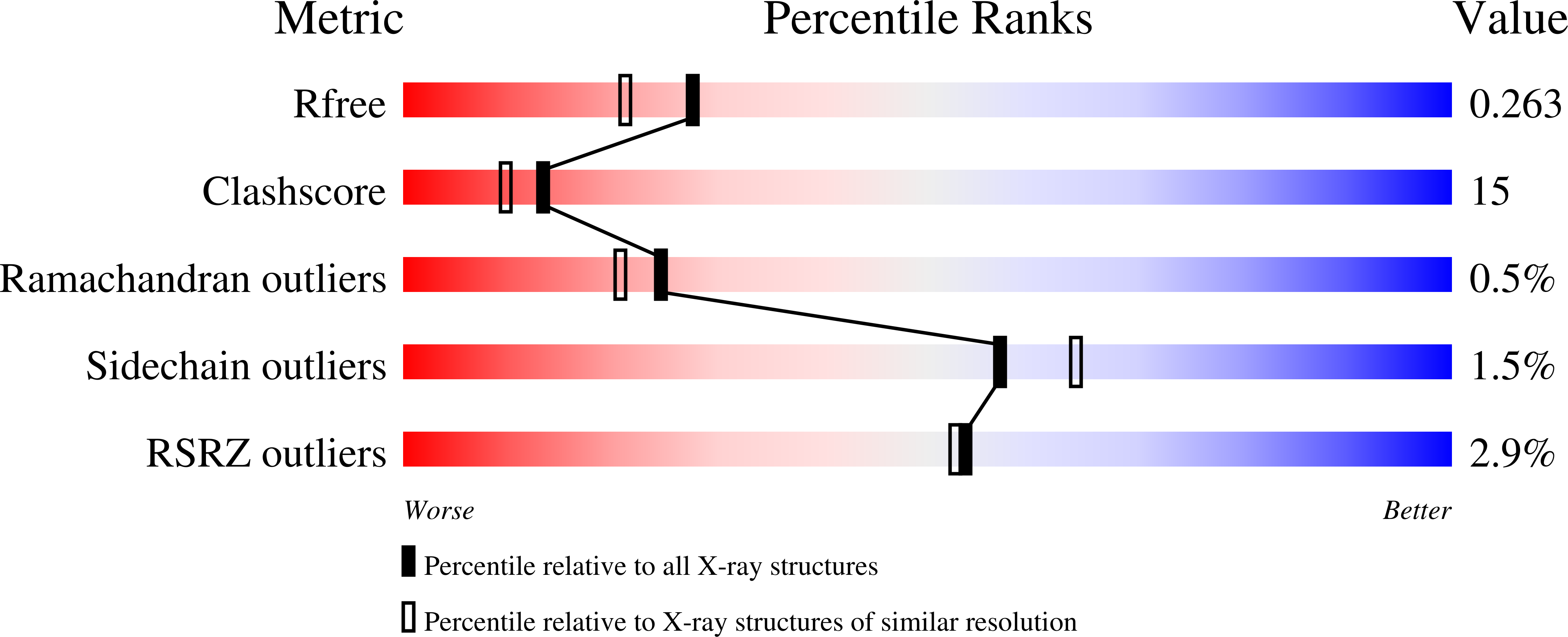
Structure analysis Details
Assembly composition:
hetero octamer (preferred)
Assembly name:
Cellular tumor antigen p53 and DNA (preferred)
PDBe Complex ID:
PDB-CPX-116221 (preferred)
Entry contents:
1 distinct polypeptide molecule
2 distinct DNA molecules
2 distinct DNA molecules
Macromolecules (3 distinct):
Cellular tumor antigen p53
Molecule details ›
Chains: A, B
Length: 197 amino acids
Theoretical weight: 22.38 KDa
Source organism: Mus musculus
Expression system: Escherichia coli
UniProt:
Sequence domains: P53 DNA-binding domain
Structure domains: Immunoglobulin-like
Length: 197 amino acids
Theoretical weight: 22.38 KDa
Source organism: Mus musculus
Expression system: Escherichia coli
UniProt:
- Canonical:
 P02340 (Residues: 96-292; Coverage: 51%)
Sequence domains: P53 DNA-binding domain
Structure domains: Immunoglobulin-like


