
ETHER</span>;</li> <li class='image_legend_li'>1 copy of <span class='highlight'>water</span>.</li></ul>")
ETHER</span>;</li> <li class='image_legend_li'>1 copy of <span class='highlight'>water</span>.</li></ul>")
ETHER</span>;</li> <li class='image_legend_li'>1 copy of <span class='highlight'>water</span>.</li></ul>")
Function and Biology Details
Biochemical function:
Biological process:
Cellular component:
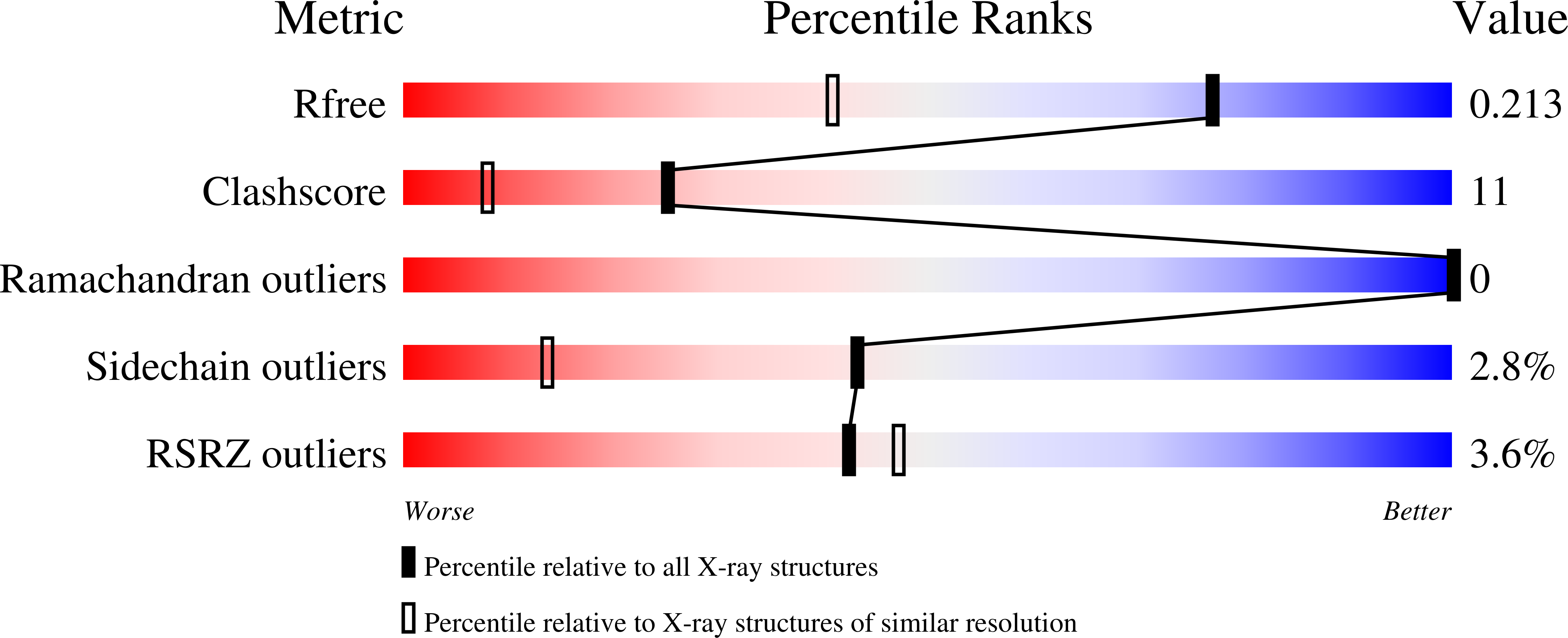
Structure analysis Details
Assembly composition:
monomeric (preferred)
Assembly name:
Cellular retinoic acid-binding protein 2 (preferred)
PDBe Complex ID:
PDB-CPX-151430 (preferred)
Entry contents:
1 distinct polypeptide molecule
Macromolecule:
Cellular retinoic acid-binding protein 2
Molecule details ›
Chains: A, B
Length: 137 amino acids
Theoretical weight: 15.49 KDa
Source organism: Homo sapiens
Expression system: Escherichia coli
UniProt:
Sequence domains: Lipocalin / cytosolic fatty-acid binding protein family
Structure domains: Lipocalin
Length: 137 amino acids
Theoretical weight: 15.49 KDa
Source organism: Homo sapiens
Expression system: Escherichia coli
UniProt:
- Canonical:
 P29373 (Residues: 2-138; Coverage: 99%)
Sequence domains: Lipocalin / cytosolic fatty-acid binding protein family
Structure domains: Lipocalin


